
Descending aorta
Citation, DOI, disclosures and article data
At the time the article was created Omar Bashir had no recorded disclosures.
View Omar Bashir's current disclosuresAt the time the article was last revised Jeremy Jones had no financial relationships to ineligible companies to disclose.
View Jeremy Jones's current disclosures- Descending thoracic aorta
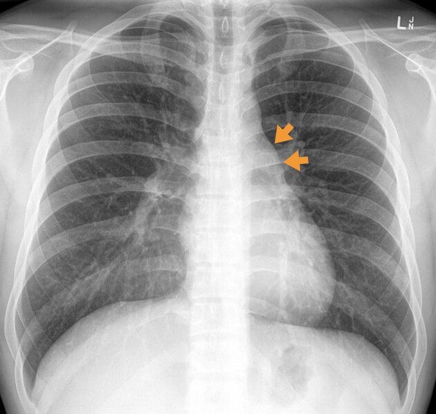
The descending aorta is the continuation of the aortic arch in the posterior mediastinum.
Gross anatomy
The descending aorta commences at the level of the fourth thoracic vertebra body, on its left, in the plane of Ludwig as the continuation of the aortic arch. It descends in the posterior mediastinum initially on the left of the thoracic vertebral bodies, but then in the midline. It exits the thorax by continuing as the abdominal aorta at the aortic diaphragmatic hiatus at the level of the twelfth thoracic vertebra 1.
Branches
pericardial branches
mediastinal arteries
esophageal arteries
Relations
Anteriorly (from superior to inferior):
left main bronchus and the hilum of the left lung
Posteriorly (from superior to inferior):
necks of the ribs of left 5 and 6th ribs
To the right (from superior to inferior):
right pleura and lung
upper esophagus
To the left (from superior to inferior):
left pleura and lung
lower esophagus
Quiz questions
References
- 1. Susan Standring. Gray's Anatomy. (2008) ISBN: 9780443066849 - Google Books
- 2. Richard S. Snell. Clinical Anatomy. (2004) ISBN: 9780781743167 - Google Books
- 3. Keith L. Moore, Arthur F. Dalley, A. M. R. Agur. Clinically Oriented Anatomy. (2013) ISBN: 9781451119459 - Google Books
- 4. Last, R. J., McMinn, R. M. H.. Last's Anatomy, Regional and Applied. (1994) ISBN: 044304662X - Google Books
- 5. Paul Butler, Adam Mitchell, Jeremiah C. Healy et al. Applied Radiological Anatomy. (2012) ISBN: 9780521766661 - Google Books
- 6. Robert H. Whitaker, Neil R. Borley. Instant Anatomy. (2000) ISBN: 9780632054039 - Google Books
Incoming Links
- Aortic valve regurgitation
- Point-of-care ultrasound (curriculum)
- Thoracic duct
- Penetrating atherosclerotic ulcer
- Thoracic aortic aneurysm
- Posterior intercostal arteries
- Ascending aortic aneurysm
- Double aortic arch
- Subcostal artery
- Minimal aortic injury
- Thoracic aorta
- Mediastinum (ITMIG classification)
- Left lower lobe consolidation
- Left paramediastinal catheter position (differential)
- Right-sided aortic arch
- Acute aortic syndrome
- Aorta
- Figure 3 sign (aortic coarctation)
- Artery of Adamkiewicz
- Aortic arch
- Abdominal aortic aneurysm
- Incomplete double aortic arch
- Patent ductus arteriosus clipping and coarctation of the aorta
- Double aortic arch
- Double outlet right ventricle
- Aortic dissection with renal ischaemia
- Double aortic arch
- Coarctation of the aorta and patent ductus arteriosus
- Cardiac MRI - axial (anatomy quiz)
- Extrapleural fat sign
- Transcatheter closure of patent ductus arteriosus
- Transcatheter closure of patent ductus arteriosus
- Double aortic arch
- Interrupted aortic arch - type A1
- Interrupted aortic arch (type A1) with Eisenmenger syndrome
- Pulmonary atresia with ventricular septal defect and situs inversus totalis
Related articles: Anatomy: Thoracic
- thoracic skeleton[+][+]
- thoracic cage
- thoracic spine
- articulations
- muscles of the thorax[+][+]
- diaphragm
- intercostal space
- intercostal muscles
- variant anatomy
- spaces of the thorax[+][+]
- thoracic viscera[+][+]
- lower respiratory tract
-
heart
- cardiac chambers
- heart valves
- cardiac fibrous skeleton
- innervation of the heart
- development of the heart
- cardiac wall
-
pericardium
- epicardium
- epicardial fat pad
- pericardial space
- oblique pericardial sinus
- transverse pericardial sinus
-
pericardial recesses
- aortic recesses
- pulmonic recesses
- postcaval recess
- pulmonary venous recesses
- pericardial ligaments
- myocardium
- endocardium
-
pericardium
- esophagus
- thymus
- breast
- arterial supply of the thorax
-
thoracic aorta (development)
-
ascending aorta[+][+]
-
aortic root
- aortic annulus
-
coronary arteries
- coronary arterial dominance
- myocardial segments
-
left main coronary artery (LMCA)
- ramus intermedius artery (RI)
-
circumflex artery (LCx)
- obtuse marginal branches (OM1, OM2, etc))
- Kugel's artery
-
left anterior descending artery (LAD)
- diagonal branches (D1, D2, etc)
- septal perforators (S1, S2, etc)
-
right coronary artery (RCA)
- conus artery
- sinoatrial nodal artery
- acute marginal branches (AM1, AM2, etc)
- inferior interventricular artery (PDA)
- posterior left ventricular artery (PLV)
- congenital anomalies
- sinotubular junction
-
aortic root
- aortic arch[+][+]
- aortic isthmus[+][+]
- descending aorta
-
ascending aorta[+][+]
- pulmonary trunk[+][+]
-
thoracic aorta (development)
- venous drainage of the thorax[+][+]
- superior vena cava (SVC)
- inferior vena cava (IVC)
-
coronary veins
-
cardiac veins which drain into the coronary sinus
- great cardiac vein
- middle cardiac vein
- small cardiac vein
- posterior vein of the left ventricle
- vein of Marshall (oblique vein of the left atrium)
- anterior cardiac veins
- venae cordis minimae (smallest cardiac veins or thebesian veins)
-
cardiac veins which drain into the coronary sinus
- pulmonary veins
- bronchial veins
- thoracoepigastric vein
- lymphatics of the thorax[+][+]
- innervation of the thorax[+][+]


 Unable to process the form. Check for errors and try again.