
Moderator band
Citation, DOI, disclosures and article data
At the time the article was created Matt A. Morgan had no recorded disclosures.
View Matt A. Morgan's current disclosuresAt the time the article was last revised Daniel J Bell had no recorded disclosures.
View Daniel J Bell's current disclosures- Septomarginal trabecula
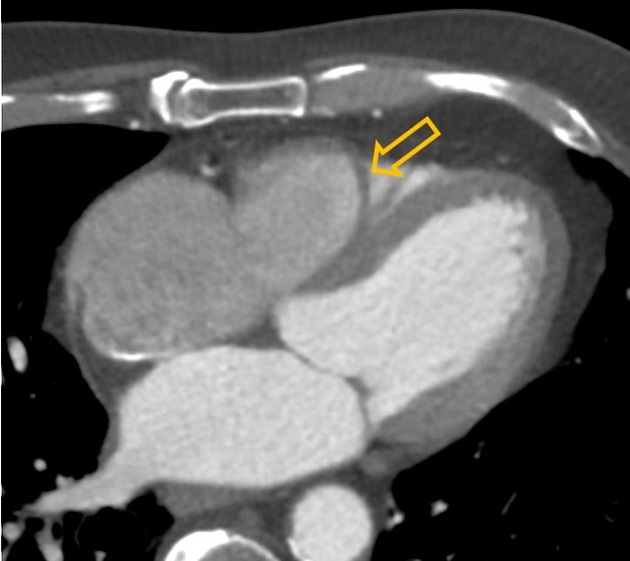
The moderator band, also called the septomarginal trabecula, is a consistent structure in the morphologic right ventricle and can be helpful as a landmark in situations where the ventricles may be ambiguous (i.e. in some forms of congenital heart disease).
The term "septomarginal" is descriptive since the muscle extends from the interventricular septum to the margin of the right ventricle, contacting/joining the base of the anterior papillary muscle.
The moderator band does not attach to the tricuspid valve, but acts as part of the electrical conduction pathway of the heart (part of the right bundle branch). Blood supply typically originates from septal perforating branches of the left anterior descending artery (LAD) which may subsequently anastomose with vessels derived from the right coronary circulation.
References
- 1. Fuster V, Walsh R, Harrington R. Hurst's the heart. McGraw-Hill Professional. ISBN:0071636463. Read it at Google Books - Find it at Amazon
- 2. Broderick LS, Brooks GN, Kuhlman JE. Anatomic pitfalls of the heart and pericardium. Radiographics. 2005;25 (2): 441-53. doi:10.1148/rg.252045075 - Pubmed citation
- 3. O'Brien JP, Srichai MB, Hecht EM et-al. Anatomy of the heart at multidetector CT: what the radiologist needs to know. Radiographics. 2007;27 (6): 1569-82. Radiographics (full text) - doi:10.1148/rg.276065747 - Pubmed citation
- 4. Prabhakar Rajiah, James MacNamara, Abhishek Chaturvedi, Ravi Ashwath, Nicholas L. Fulton, Harold Goerne. Bands in the Heart: Multimodality Imaging Review. (2019) RadioGraphics. 39 (5): 1238-1263. doi:10.1148/rg.2019180176 - Pubmed
- 5. Lee JY, Hur MS. Morphological classification of the moderator band and its relationship with the anterior papillary muscle. (2019) Anatomy & cell biology. 52 (1): 38-42. doi:10.5115/acb.2019.52.1.38 - Pubmed
- 6. Terpenning S, White CS. Imaging pitfalls, normal anatomy, and anatomical variants that can simulate disease on cardiac imaging as demonstrated on multidetector computed tomography. (2015) Acta radiologica short reports. 4 (1): 2047981614562443. doi:10.1177/2047981614562443 - Pubmed
- 7. Reig J, Albertí N, Petit M. Arterial vascularization of the human moderator band: an analysis of this structure's role as a collateral circulation route. (2000) Clinical anatomy (New York, N.Y.). 13 (4): 244-50. doi:10.1002/1098-2353(2000)13:4<244::AID-CA3>3.0.CO;2-H - Pubmed
Incoming Links
Related articles: Anatomy: Thoracic
- thoracic skeleton[+][+]
- thoracic cage
- thoracic spine
- articulations
- muscles of the thorax[+][+]
- diaphragm
- intercostal space
- intercostal muscles
- variant anatomy
- spaces of the thorax[+][+]
- thoracic viscera
- lower respiratory tract[+][+]
-
heart
- cardiac chambers
- heart valves[+][+]
- cardiac fibrous skeleton
- innervation of the heart
- development of the heart[+][+]
- cardiac wall[+][+]
-
pericardium
- epicardium
- epicardial fat pad
- pericardial space
- oblique pericardial sinus
- transverse pericardial sinus
-
pericardial recesses
- aortic recesses
- pulmonic recesses
- postcaval recess
- pulmonary venous recesses
- pericardial ligaments
- myocardium
- endocardium
-
pericardium
- esophagus[+][+]
- thymus[+][+]
- breast[+][+]
- arterial supply of the thorax[+][+]
-
thoracic aorta (development)
-
ascending aorta
-
aortic root
- aortic annulus
-
coronary arteries
- coronary arterial dominance
- myocardial segments
-
left main coronary artery (LMCA)
- ramus intermedius artery (RI)
-
circumflex artery (LCx)
- obtuse marginal branches (OM1, OM2, etc))
- Kugel's artery
-
left anterior descending artery (LAD)
- diagonal branches (D1, D2, etc)
- septal perforators (S1, S2, etc)
-
right coronary artery (RCA)
- conus artery
- sinoatrial nodal artery
- acute marginal branches (AM1, AM2, etc)
- inferior interventricular artery (PDA)
- posterior left ventricular artery (PLV)
- congenital anomalies
- sinotubular junction
-
aortic root
- aortic arch
- aortic isthmus
- descending aorta
-
ascending aorta
- pulmonary trunk
-
thoracic aorta (development)
- venous drainage of the thorax[+][+]
- superior vena cava (SVC)
- inferior vena cava (IVC)
-
coronary veins
-
cardiac veins which drain into the coronary sinus
- great cardiac vein
- middle cardiac vein
- small cardiac vein
- posterior vein of the left ventricle
- vein of Marshall (oblique vein of the left atrium)
- anterior cardiac veins
- venae cordis minimae (smallest cardiac veins or thebesian veins)
-
cardiac veins which drain into the coronary sinus
- pulmonary veins
- bronchial veins
- thoracoepigastric vein
- lymphatics of the thorax[+][+]
- innervation of the thorax[+][+]


 Unable to process the form. Check for errors and try again.