
The pericardium is a conical, flask-like, fibroserous sac which contains the heart and the roots of the great vessels and defines the middle mediastinum.
On this page:
Gross anatomy
The pericardium is made of two sacs in one. The outer sac is the fibrous pericardium and the inner sac is the double-layered serous pericardium. Layers of serous pericardium are divided by the pericardial space, which only contains 15-50 mL of serous fluid 10. Each layer has quite different structure and functions (from external to internal):
fibrous pericardium: tough connective tissue continuous with and bound to the central tendon of the diaphragm (pericardiophrenic ligament), the roots of the great vessels, the pretracheal layer of the deep cervical fascia and the sternum via the superior (to manubrium) and inferior sternopericardial ligaments (to xiphoid process)
-
serous pericardium: composed of a single layer of flattened mesothelial cells reflected on itself at the root of the great vessels to form a closed sac. The two layers are:
parietal serous pericardium: lines the deep surface of the fibrous pericardium and is inseparable from it
visceral serous pericardium: covers heart and great vessels forming the epicardium. Some fat exists between the epicardium and myocardium and it increases with age. The amount of fat may become more extensive at the anterior and lateral cardiophrenic angles, where it is known as pericardial fat pad 10.
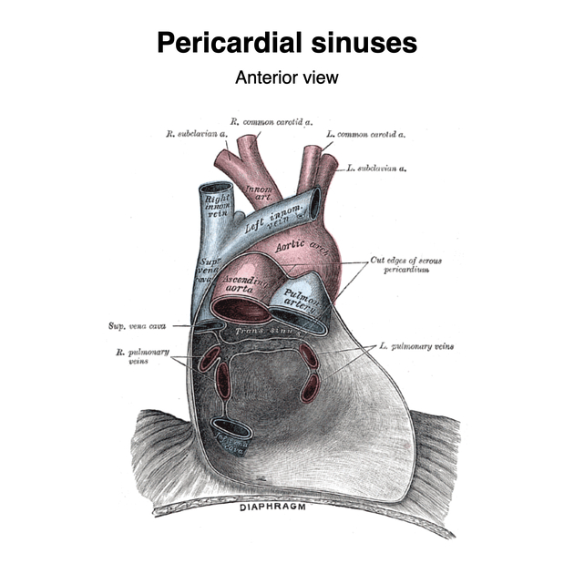
The serous pericardium is invaginated by the heart and great vessels forming two sinuses:
oblique pericardial sinus: blind-ending, inverted U-shaped space posterior to the left atrium which is continuous with the pericardial cavity proper inferiorly. It is bound by the pericardial reflection around the superior and inferior vena cavae and the four pulmonary veins. This sinus may be mistaken for esophageal masses or bronchogenic cysts 3
transverse pericardial sinus: tunnel-like communication between the right and left sides of the pericardial cavity proper. It lies posterior to the root of the aortic arch and pulmonary trunk, and superior to the left atrium. Several pericardial recesses are created by the transverse pericardial sinus which may be mistaken for dissection or lymphadenopathy 3
Adjacent to these sinuses, there may be one or several pericardial recesses:
-
aortic recesses - arise from the transverse sinus
superior aortic recess: from its mouth located inferiorly, it ascends posterior to and then to the right of the ascending aorta and ends at the level of the sternal angle
inferior aortic recess: diverticulum descending from the superiorly located mouth to run between the lower ascending part of the aorta and the right atrium
-
pulmonic recesses - arise from transverse sinus
right pulmonic recess: posterior to the right pulmonary artery and anterior to the esophagus
left pulmonic recess: bounded superiorly by the left pulmonary artery, inferiorly by the left superior pulmonary vein and medially by the ligament of Marshall
postcaval recess: posterior to the superior vena cava, superior to the right superior pulmonary vein and inferior to the right pulmonary artery, with a mouth that opens superolaterally to the right
posterior pericardial recess: arises superiorly from oblique sinus, posterior to the right pulmonary artery and medial to the bronchus intermedius
-
pulmonary venous recesses - arise from the pericardial cavity proper
-
right pulmonary venous recess and left pulmonary venous recess
project medially and upward on the back of the left atrium between the superior and inferior pulmonary veins on each side, indenting the side walls of the oblique sinus
-
Relations
anteriorly: body of the sternum, cartilages of left third to seventh ribs, pleura and lungs, and thymus (in children)
posteriorly: esophagus, descending thoracic aorta, pleura and lungs
laterally: pleura and lungs, phrenic nerves
inferiorly: blends with central tendon of diaphragm
Arterial supply
internal thoracic artery and its pericardiophrenic and musculophrenic branches
branches from the descending thoracic aorta
Venous drainage
Innervation
phrenic nerves: to fibrous and parietal serous layers
sympathetic trunks: for pain, muscles and vessels of heart
Lymphatic drainage
tracheobronchial nodes
prepericardial nodes 9
Variant anatomy
Radiographic appearance
CT
pericardium appears as a thin high-density line between the lower-density mediastinal and epicardial fat 10
low density pericardial recesses and sinuses between the great vessels should not be mistakened as lymph nodes 10
fibrous and serous pericardium cannot be delineated and the upper limits of normal for pericardial thickness is 2 mm 2
often not imaged over the left ventricle 3
MRI
T1 and T2: appears as a low signal rim between the higher-signal mediastinal and epicardial fat 2,3
Related pathology
pericardial fat tag sign of pneumothorax on supine CXR


 Unable to process the form. Check for errors and try again.