
Superior aortic recess
Last revised by Jose Antonio Habana
on 16 Oct 2024
Citation, DOI, disclosures and article data
Citation:
Tromp D, Habana J, Campos A, et al. Superior aortic recess. Reference article, Radiopaedia.org (Accessed on 25 Mar 2025) https://doi.org/10.53347/rID-47077
Permalink:
rID:
47077
Article created:
30 Jul 2016,
Dirk Tromp
Disclosures:
At the time the article was created Dirk Tromp had no recorded disclosures.
View Dirk Tromp's current disclosures
Last revised:
16 Oct 2024,
Jose Antonio Habana
Disclosures:
At the time the article was last revised Jose Antonio Habana had no financial relationships to ineligible companies to disclose.
View Jose Antonio Habana's current disclosures
Revisions:
15 times, by
10 contributors -
see full revision history and disclosures
Systems:
Sections:
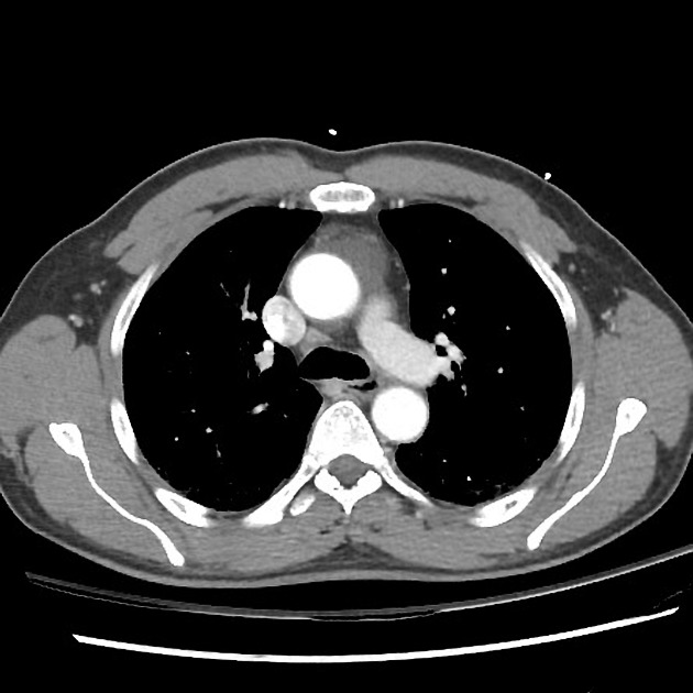
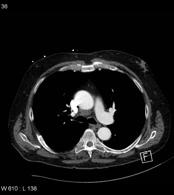
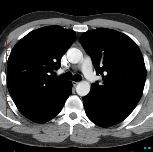
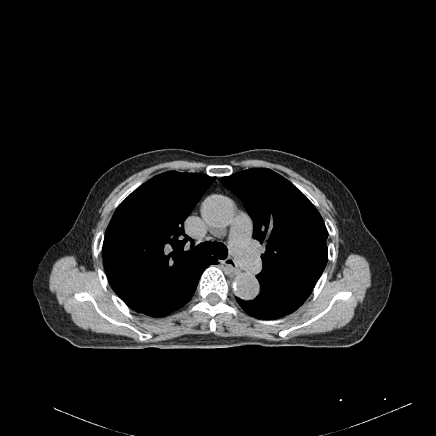
The superior aortic recess is one of the pericardial recesses forming a small space within the pericardium, which arises from the superior margin of the transverse pericardial sinus and surrounds the root of the ascending aorta.
Its components are variable and may be further subdivided into:
- anterior superior aortic recess
- right superior aortic recess
- posterior superior aortic recess (also called the superior sinus or superior pericardial recess)
It may mimic mediastinal lymphadenopathy or a bronchogenic cyst.
References
- 1. Groell R, Schaffler GJ, Rienmueller R. Pericardial sinuses and recesses: findings at electrocardiographically triggered electron-beam CT. Radiology. 1999;212 (1): 69-73. doi:10.1148/radiology.212.1.r99jl0969 - Pubmed citation
- 2. Truong MT, Erasmus JJ, Gladish GW et-al. Anatomy of pericardial recesses on multidetector CT: implications for oncologic imaging. AJR Am J Roentgenol. 2003;181 (4): 1109-13. doi:10.2214/ajr.181.4.1811109 - Pubmed citation
- 3. Oyama N, Oyama N, Komuro K et-al. Computed tomography and magnetic resonance imaging of the pericardium: anatomy and pathology. Magnetic resonance in medical sciences: MRMS: an official journal of Japan Society of Magnetic Resonance in Medicine. 3 (3): 145-52. Pubmed
- 4. O'Leary SM, Williams PL, Williams MP et-al. Imaging the pericardium: appearances on ECG-gated 64-detector row cardiac computed tomography. Br J Radiol. 2010;83 (987): 194-205. Br J Radiol (full text) - doi:10.1259/bjr/55699491 - Free text at pubmed - Pubmed citation
- 5. Wang ZJ, Reddy GP, Gotway MB et-al. CT and MR imaging of pericardial disease. Radiographics : a review publication of the Radiological Society of North America, Inc. 23 Spec No: S167-80. doi:10.1148/rg.23si035504 - Pubmed
- 6. Broderick LS, Brooks GN, Kuhlman JE. Anatomic pitfalls of the heart and pericardium. Radiographics : a review publication of the Radiological Society of North America, Inc. 25 (2): 441-53. doi:10.1148/rg.252045075 - Pubmed
Incoming Links
Related articles: Anatomy: Thoracic
- thoracic skeleton[+][+]
- thoracic cage
- thoracic spine
- articulations
- muscles of the thorax[+][+]
- diaphragm
- intercostal space
- intercostal muscles
- variant anatomy
- spaces of the thorax[+][+]
- thoracic viscera
- lower respiratory tract[+][+]
-
heart
- cardiac chambers[+][+]
- heart valves[+][+]
- cardiac fibrous skeleton
- innervation of the heart
- development of the heart[+][+]
- cardiac wall
-
pericardium
- epicardium
- epicardial fat pad
- pericardial space
- oblique pericardial sinus
- transverse pericardial sinus
-
pericardial recesses
- aortic recesses
- superior aortic recess
- inferior aortic recess
- pulmonic recesses[+][+]
- postcaval recess
- pulmonary venous recesses[+][+]
- aortic recesses
- pericardial ligaments
- myocardium
- endocardium
-
pericardium
- esophagus[+][+]
- thymus[+][+]
- breast[+][+]
- arterial supply of the thorax[+][+]
-
thoracic aorta (development)
-
ascending aorta
-
aortic root
- aortic annulus
-
coronary arteries
- coronary arterial dominance
- myocardial segments
-
left main coronary artery (LMCA)
- ramus intermedius artery (RI)
-
circumflex artery (LCx)
- obtuse marginal branches (OM1, OM2, etc))
- Kugel's artery
-
left anterior descending artery (LAD)
- diagonal branches (D1, D2, etc)
- septal perforators (S1, S2, etc)
-
right coronary artery (RCA)
- conus artery
- sinoatrial nodal artery
- acute marginal branches (AM1, AM2, etc)
- inferior interventricular artery (PDA)
- posterior left ventricular artery (PLV)
- congenital anomalies
- sinotubular junction
-
aortic root
- aortic arch
- aortic isthmus
- descending aorta
-
ascending aorta
- pulmonary trunk
-
thoracic aorta (development)
- venous drainage of the thorax[+][+]
- superior vena cava (SVC)
- inferior vena cava (IVC)
-
coronary veins
-
cardiac veins which drain into the coronary sinus
- great cardiac vein
- middle cardiac vein
- small cardiac vein
- posterior vein of the left ventricle
- vein of Marshall (oblique vein of the left atrium)
- anterior cardiac veins
- venae cordis minimae (smallest cardiac veins or thebesian veins)
-
cardiac veins which drain into the coronary sinus
- pulmonary veins
- bronchial veins
- thoracoepigastric vein
- lymphatics of the thorax[+][+]
- innervation of the thorax[+][+]


 Unable to process the form. Check for errors and try again.