
Ramus intermedius artery
Citation, DOI, disclosures and article data
At the time the article was created Henry Knipe had no recorded disclosures.
View Henry Knipe's current disclosuresAt the time the article was last revised Joachim Feger had no financial relationships to ineligible companies to disclose.
View Joachim Feger's current disclosures- Ramus intermedius
- Ramus medianus
- RI
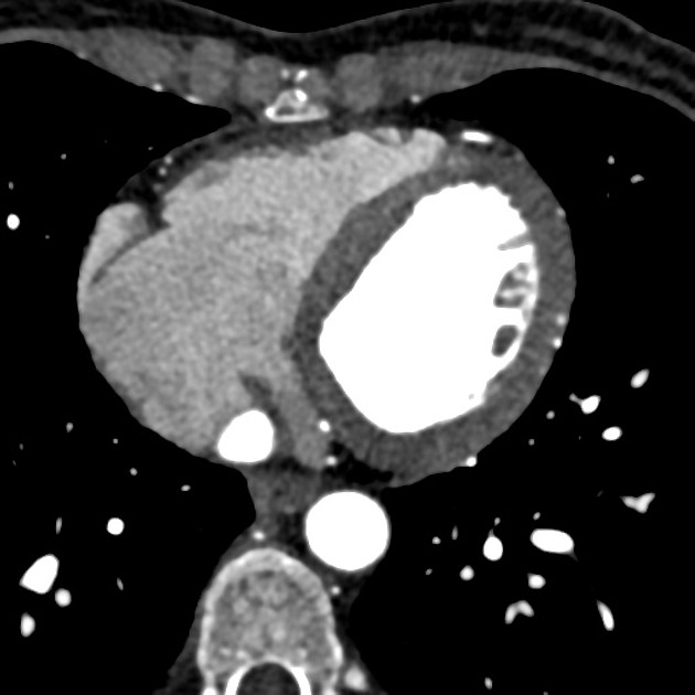
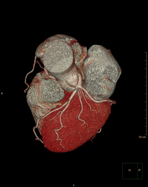
The ramus intermedius is a variant coronary artery resulting from trifurcation of the left main coronary artery 1. It is present in ~20% (range 15-30%) 2,3 of the population.
It can have a course similar to the obtuse marginal branches of the left circumflex artery or the diagonal branches of the left anterior descending artery and thus can supply either the anterior or medial aspect of the heart 1.
The peculiarity of this vessel is that it does not run along an anatomical groove and simply slides over the free surface of the left ventricle instead.
Practical points
In practice, even if there is not a true trifurcation, some cardiologists may term a prominent early branching (high origin) obtuse marginal artery a ramus branch if it supplies the territory of small diagonal branches since there is an inverse relationship between the size of the ramus intermedius and the size and distribution of the diagonal branches.
References
- 1. Kini S, Bis K, Weaver L. Normal and Variant Coronary Arterial and Venous Anatomy on High-Resolution CT Angiography. AJR Am J Roentgenol. 2007;188(6):1665-74. doi:10.2214/AJR.06.1295 - Pubmed
- 2. Koşar P, Ergun E, Oztürk C, Koşar U. Anatomic Variations and Anomalies of the Coronary Arteries: 64-Slice CT Angiographic Appearance. Diagn Interv Radiol. 2009;15(4):275-83. doi:10.4261/1305-3825.DIR.2550-09.1 - Pubmed
- 3. O'Brien J, Srichai M, Hecht E, Kim D, Jacobs J. Anatomy of the Heart at Multidetector CT: What the Radiologist Needs to Know. Radiographics. 2007;27(6):1569-82. doi:10.1148/rg.276065747 - Pubmed
- 4. Paolillo V, Gastaldo D, Vaudano G. An Unusual Course of the Ramus Intermedius: Shown by Multislice Computed Tomographic Coronary Angiography. Tex Heart Inst J. 2006;33(3):406-7. PMC1592261 - Pubmed
- 5. Paolillo V, Gastaldo D, Vaudano G. An Unusual Course of the Ramus Intermedius: Shown by Multislice Computed Tomographic Coronary Angiography. Tex Heart Inst J. 2006;33(3):406-7. PMC1592261 - Pubmed
Incoming Links
Related articles: Anatomy: Thoracic
- thoracic skeleton[+][+]
- thoracic cage
- thoracic spine
- articulations
- muscles of the thorax[+][+]
- diaphragm
- intercostal space
- intercostal muscles
- variant anatomy
- spaces of the thorax[+][+]
- thoracic viscera[+][+]
- lower respiratory tract
-
heart
- cardiac chambers
- heart valves
- cardiac fibrous skeleton
- innervation of the heart
- development of the heart
- cardiac wall
-
pericardium
- epicardium
- epicardial fat pad
- pericardial space
- oblique pericardial sinus
- transverse pericardial sinus
-
pericardial recesses
- aortic recesses
- pulmonic recesses
- postcaval recess
- pulmonary venous recesses
- pericardial ligaments
- myocardium
- endocardium
-
pericardium
- esophagus
- thymus
- breast
- arterial supply of the thorax
-
thoracic aorta (development)
-
ascending aorta
-
aortic root
- aortic annulus
-
coronary arteries
- coronary arterial dominance
- myocardial segments
-
left main coronary artery (LMCA)
- ramus intermedius artery (RI)
-
circumflex artery (LCx)[+][+]
- obtuse marginal branches (OM1, OM2, etc))
- Kugel's artery
-
left anterior descending artery (LAD)[+][+]
- diagonal branches (D1, D2, etc)
- septal perforators (S1, S2, etc)
-
right coronary artery (RCA)[+][+]
- conus artery
- sinoatrial nodal artery
- acute marginal branches (AM1, AM2, etc)
- inferior interventricular artery (PDA)
- posterior left ventricular artery (PLV)
- congenital anomalies
- sinotubular junction
-
aortic root
- aortic arch[+][+]
- aortic isthmus[+][+]
- descending aorta[+][+]
-
ascending aorta
- pulmonary trunk[+][+]
-
thoracic aorta (development)
- venous drainage of the thorax[+][+]
- superior vena cava (SVC)
- inferior vena cava (IVC)
-
coronary veins
-
cardiac veins which drain into the coronary sinus
- great cardiac vein
- middle cardiac vein
- small cardiac vein
- posterior vein of the left ventricle
- vein of Marshall (oblique vein of the left atrium)
- anterior cardiac veins
- venae cordis minimae (smallest cardiac veins or thebesian veins)
-
cardiac veins which drain into the coronary sinus
- pulmonary veins
- bronchial veins
- thoracoepigastric vein
- lymphatics of the thorax[+][+]
- innervation of the thorax[+][+]


 Unable to process the form. Check for errors and try again.