
Intercostal muscles
Citation, DOI, disclosures and article data
Citation:
Knipe H, Vadera S, Kang O, et al. Intercostal muscles. Reference article, Radiopaedia.org (Accessed on 08 Mar 2025) https://doi.org/10.53347/rID-26642
Permalink:
rID:
26642
Article created:
Disclosures:
At the time the article was created Henry Knipe had no recorded disclosures.
View Henry Knipe's current disclosures
Last revised:
Disclosures:
At the time the article was last revised Sonam Vadera had no recorded disclosures.
View Sonam Vadera's current disclosures
Revisions:
6 times, by
5 contributors -
see full revision history and disclosures
Systems:
Sections:
Synonyms:
- Intercostal muscle
- Intercostal muscle group
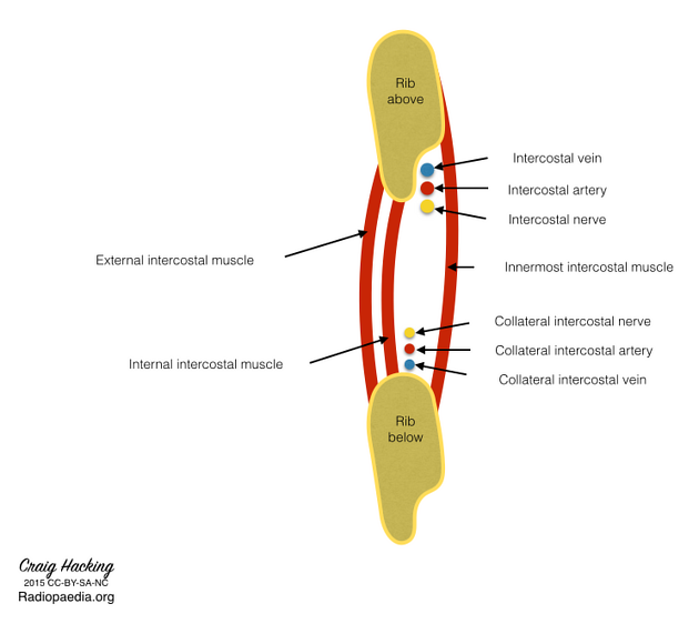
The intercostal muscles are an important group of muscles in the intercostal spaces (between the ribs) that contract during respiration. Three muscles are classically described, from superficial to deep:
The following two muscles can also be considered part of this group:
References
- 1. Platzer W. Color Atlas of Human Anatomy locomotor system, Vol. 1. Thieme. ISBN:313533306X. Read it at Google Books - Find it at Amazon
- 2. Snell RS. Clinical Anatomy by Regions. Lippincott Williams & Wilkins. ISBN:160913446X. Read it at Google Books - Find it at Amazon
- 3. Kaiser L, Singhal S. Surgical foundations. Mosby. ISBN:0815126131. Read it at Google Books - Find it at Amazon
- 4. Last's anatomy, regional and applied. Churchill Livingstone. ISBN:044304662X. Read it at Google Books - Find it at Amazon
- 5. Moore KL, Agur AMR, Dalley AF. Clinically oriented anatomy. LWW. ISBN:1451119453. Read it at Google Books - Find it at Amazon
Incoming Links
Articles:
Related articles: Anatomy: Thoracic
- thoracic skeleton[+][+]
- thoracic cage
- thoracic spine
- articulations
- muscles of the thorax
- spaces of the thorax[+][+]
- thoracic viscera[+][+]
- lower respiratory tract
-
heart
- cardiac chambers
- heart valves
- cardiac fibrous skeleton
- innervation of the heart
- development of the heart
- cardiac wall
-
pericardium
- epicardium
- epicardial fat pad
- pericardial space
- oblique pericardial sinus
- transverse pericardial sinus
-
pericardial recesses
- aortic recesses
- pulmonic recesses
- postcaval recess
- pulmonary venous recesses
- pericardial ligaments
- myocardium
- endocardium
-
pericardium
- esophagus
- thymus
- breast
- arterial supply of the thorax[+][+]
-
thoracic aorta (development)
-
ascending aorta
-
aortic root
- aortic annulus
-
coronary arteries
- coronary arterial dominance
- myocardial segments
-
left main coronary artery (LMCA)
- ramus intermedius artery (RI)
-
circumflex artery (LCx)
- obtuse marginal branches (OM1, OM2, etc))
- Kugel's artery
-
left anterior descending artery (LAD)
- diagonal branches (D1, D2, etc)
- septal perforators (S1, S2, etc)
-
right coronary artery (RCA)
- conus artery
- sinoatrial nodal artery
- acute marginal branches (AM1, AM2, etc)
- inferior interventricular artery (PDA)
- posterior left ventricular artery (PLV)
- congenital anomalies
- sinotubular junction
-
aortic root
- aortic arch
- aortic isthmus
- descending aorta
-
ascending aorta
- pulmonary trunk
-
thoracic aorta (development)
- venous drainage of the thorax[+][+]
- superior vena cava (SVC)
- inferior vena cava (IVC)
-
coronary veins
-
cardiac veins which drain into the coronary sinus
- great cardiac vein
- middle cardiac vein
- small cardiac vein
- posterior vein of the left ventricle
- vein of Marshall (oblique vein of the left atrium)
- anterior cardiac veins
- venae cordis minimae (smallest cardiac veins or thebesian veins)
-
cardiac veins which drain into the coronary sinus
- pulmonary veins
- bronchial veins
- thoracoepigastric vein
- lymphatics of the thorax[+][+]
- innervation of the thorax[+][+]


 Unable to process the form. Check for errors and try again.