
Right lung
Citation, DOI, disclosures and article data
Citation:
Hacking C, Skalski M, Right lung. Reference article, Radiopaedia.org (Accessed on 02 Mar 2025) https://doi.org/10.53347/rID-38934
Permalink:
rID:
38934
Article created:
Disclosures:
At the time the article was created Craig Hacking had no recorded disclosures.
View Craig Hacking's current disclosures
Last revised:
Disclosures:
At the time the article was last revised Craig Hacking had no recorded disclosures.
View Craig Hacking's current disclosures
Revisions:
5 times, by
2 contributors -
see full revision history and disclosures
Systems:
Sections:
The right lung is one of two lungs, located in the right hemithorax on the right of the heart and mediastinum.
There are a few differences between the two lungs:
- The right lung is larger in volume than the left lung, with a larger transverse dimension (due to the heart on the left) but a shorter longitudinal dimension (due to the liver causing the right hemidiaphragm to be higher than the left).
- The right main bronchus also differs to the left main bronchus, as it is longer, larger caliber and more vertical.
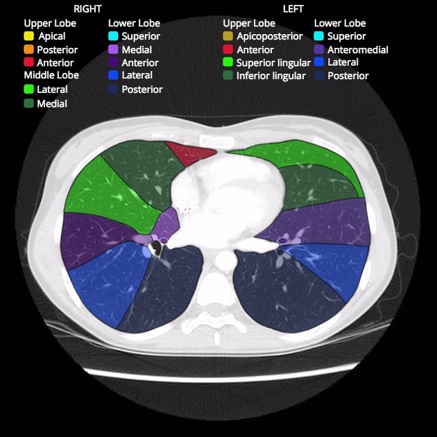
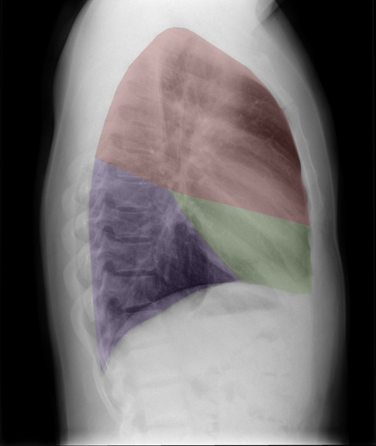
- The right lung has 2 fissures: the oblique and horizontal fissures.
- The right lung contains 3 lobes and 10 bronchopulmonary segments:
For a detailed description of lung anatomy and function, see the lung article here.
References
- 1. Moore KL, Agur AMR, Dalley AF. Clinically oriented anatomy. LWW. ISBN:1451119453. Read it at Google Books - Find it at Amazon
Incoming Links
Articles:
Cases:
Related articles: Anatomy: Thoracic
- thoracic skeleton[+][+]
- thoracic cage
- thoracic spine
- articulations
- muscles of the thorax[+][+]
- diaphragm
- intercostal space
- intercostal muscles
- variant anatomy
- spaces of the thorax[+][+]
- thoracic viscera
-
lower respiratory tract
- tracheobronchial tree[+][+]
- lungs
-
heart[+][+]
- cardiac chambers
- heart valves
- cardiac fibrous skeleton
- innervation of the heart
- development of the heart
- cardiac wall
-
pericardium
- epicardium
- epicardial fat pad
- pericardial space
- oblique pericardial sinus
- transverse pericardial sinus
-
pericardial recesses
- aortic recesses
- pulmonic recesses
- postcaval recess
- pulmonary venous recesses
- pericardial ligaments
- myocardium
- endocardium
-
pericardium
- esophagus[+][+]
- thymus[+][+]
- breast[+][+]
-
lower respiratory tract
- arterial supply of the thorax[+][+]
-
thoracic aorta (development)
-
ascending aorta
-
aortic root
- aortic annulus
-
coronary arteries
- coronary arterial dominance
- myocardial segments
-
left main coronary artery (LMCA)
- ramus intermedius artery (RI)
-
circumflex artery (LCx)
- obtuse marginal branches (OM1, OM2, etc))
- Kugel's artery
-
left anterior descending artery (LAD)
- diagonal branches (D1, D2, etc)
- septal perforators (S1, S2, etc)
-
right coronary artery (RCA)
- conus artery
- sinoatrial nodal artery
- acute marginal branches (AM1, AM2, etc)
- inferior interventricular artery (PDA)
- posterior left ventricular artery (PLV)
- congenital anomalies
- sinotubular junction
-
aortic root
- aortic arch
- aortic isthmus
- descending aorta
-
ascending aorta
- pulmonary trunk
-
thoracic aorta (development)
- venous drainage of the thorax[+][+]
- superior vena cava (SVC)
- inferior vena cava (IVC)
-
coronary veins
-
cardiac veins which drain into the coronary sinus
- great cardiac vein
- middle cardiac vein
- small cardiac vein
- posterior vein of the left ventricle
- vein of Marshall (oblique vein of the left atrium)
- anterior cardiac veins
- venae cordis minimae (smallest cardiac veins or thebesian veins)
-
cardiac veins which drain into the coronary sinus
- pulmonary veins
- bronchial veins
- thoracoepigastric vein
- lymphatics of the thorax[+][+]
- innervation of the thorax[+][+]


 Unable to process the form. Check for errors and try again.