
Left ventricular outflow tract
Citation, DOI, disclosures and article data
Citation:
Weerakkody Y, Hacking C, Carroll D, et al. Left ventricular outflow tract. Reference article, Radiopaedia.org (Accessed on 21 Feb 2025) https://doi.org/10.53347/rID-57059
Permalink:
rID:
57059
Article created:
Disclosures:
At the time the article was created Yuranga Weerakkody had no recorded disclosures.
View Yuranga Weerakkody's current disclosures
Last revised:
Disclosures:
At the time the article was last revised Craig Hacking had the following disclosures:
- Philips Australia, Paid speaker at Philips Spectral CT events (ongoing)
These were assessed during peer review and were determined to not be relevant to the changes that were made.
View Craig Hacking's current disclosures
Revisions:
7 times, by
4 contributors -
see full revision history and disclosures
Systems:
Sections:
Tags:
Synonyms:
- Left ventricular outflow tract (LVOT)
- LVOT
- Aortic vestibule
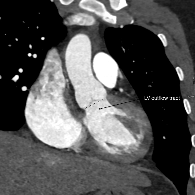
The left ventricular outflow tract (LVOT) (also sometimes called the aortic vestibule) is considered to represent the region of the left ventricle that lies between the anterior cusp of the mitral valve and the ventricular septum. It directs blood towards the aortic annulus and through the aortic valve during systole. Its dimensions are often recorded in TAVI workup studies.
Related pathology
See also
References
- 1. Renapurkar RD, El-Sherief AH, Prieto L, Kapadia SR, Schoenhagen P. Transcatheter structural cardiac intervention: a radiology perspective. AJR. American journal of roentgenology. 204 (6): W648-62. doi:10.2214/AJR.14.12571 - Pubmed
- 2. Anatomy of left ventricular outflow tract. Heart. 41 (3): 263. doi:10.1136/hrt.41.3.263 - Pubmed
- 3. Shiokawa Y, Becker AE. The surgical anatomy of the left ventricular outflow tract in hearts with ventricular septal defect and aortic arch obstruction. The Annals of thoracic surgery. 65 (5): 1381-7. Pubmed
Incoming Links
Articles:
- Left ventricular outflow tract obstruction in echocardiography (differential)
- Approach to shock (echocardiography)
- Aortic valve stenosis
- Stroke volume
- Prosthetic heart valve
- M-mode (ultrasound)
- Transcatheter mitral valve intervention
- Arterial switch procedure
- Second trimester ultrasound scan
- Interventricular septum
- Medical abbreviations and acronyms (L)
- Axenfeld-Rieger syndrome
- Left ventricular outflow tract view (fetal echocardiogram)
- Transcatheter aortic valve implantation (TAVI)
- Non-compaction of the left ventricle
- Cardiovascular shunts
Related articles: Anatomy: Thoracic
- thoracic skeleton[+][+]
- thoracic cage
- thoracic spine
- articulations
- muscles of the thorax[+][+]
- diaphragm
- intercostal space
- intercostal muscles
- variant anatomy
- spaces of the thorax[+][+]
- thoracic viscera
- lower respiratory tract[+][+]
-
heart
- cardiac chambers
- heart valves[+][+]
- cardiac fibrous skeleton
- innervation of the heart
- development of the heart[+][+]
- cardiac wall[+][+]
-
pericardium
- epicardium
- epicardial fat pad
- pericardial space
- oblique pericardial sinus
- transverse pericardial sinus
-
pericardial recesses
- aortic recesses
- pulmonic recesses
- postcaval recess
- pulmonary venous recesses
- pericardial ligaments
- myocardium
- endocardium
-
pericardium
- esophagus[+][+]
- thymus[+][+]
- breast[+][+]
- arterial supply of the thorax[+][+]
-
thoracic aorta (development)
-
ascending aorta
-
aortic root
- aortic annulus
-
coronary arteries
- coronary arterial dominance
- myocardial segments
-
left main coronary artery (LMCA)
- ramus intermedius artery (RI)
-
circumflex artery (LCx)
- obtuse marginal branches (OM1, OM2, etc))
- Kugel's artery
-
left anterior descending artery (LAD)
- diagonal branches (D1, D2, etc)
- septal perforators (S1, S2, etc)
-
right coronary artery (RCA)
- conus artery
- sinoatrial nodal artery
- acute marginal branches (AM1, AM2, etc)
- inferior interventricular artery (PDA)
- posterior left ventricular artery (PLV)
- congenital anomalies
- sinotubular junction
-
aortic root
- aortic arch
- aortic isthmus
- descending aorta
-
ascending aorta
- pulmonary trunk
-
thoracic aorta (development)
- venous drainage of the thorax[+][+]
- superior vena cava (SVC)
- inferior vena cava (IVC)
-
coronary veins
-
cardiac veins which drain into the coronary sinus
- great cardiac vein
- middle cardiac vein
- small cardiac vein
- posterior vein of the left ventricle
- vein of Marshall (oblique vein of the left atrium)
- anterior cardiac veins
- venae cordis minimae (smallest cardiac veins or thebesian veins)
-
cardiac veins which drain into the coronary sinus
- pulmonary veins
- bronchial veins
- thoracoepigastric vein
- lymphatics of the thorax[+][+]
- innervation of the thorax[+][+]


 Unable to process the form. Check for errors and try again.