
The superior thoracic aperture, also known as the thoracic inlet or outlet, connects the root of the neck with the thorax.
On this page:
Article:
Images:
Images:
Gross anatomy
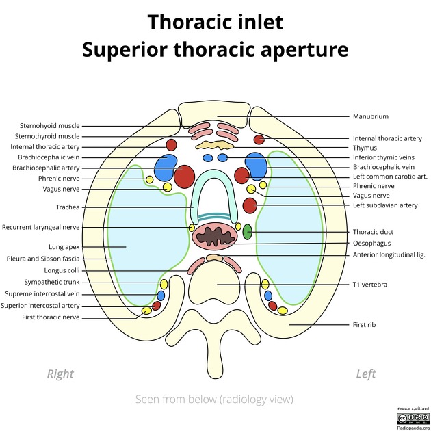
The superior thoracic aperture is kidney-shaped and lies in an oblique transverse plane, tilted anteroinferiorly to posterosuperiorly. It is roughly 10 cm in transverse dimension and 5 cm in AP dimension.
Boundaries
- posteriorly: T1 vertebral body and costovertebral joints
- laterally: first ribs and their costal cartilages
- anteriorly: superior border of the manubrium
Contents
The list of structures that pass through the superior thoracic aperture is long and can be divided into five groups: midline, bilateral, posteriorly, and asymmetric left and right.
- midline from anterior to posterior
- sternohyoid and sternothyroid muscles
- thymic remnants
- inferior thyroid veins
- trachea
- tracheo-esophageal sulcus containing recurrent laryngeal nerves
- esophagus
- thoracic duct displaced to the left
- longus colli muscles
- anterior longitudinal ligament
- laterally on both sides
- posteriorly from medial to lateral
- sympathetic trunk
- supreme intercostal vein
- superior intercostal artery
- ventral ramus of the first thoracic nerve
- on the left:
- left common carotid artery
- left subclavian artery
- left vagus nerve, between left common carotid and left subclavian arteries
- left brachiocephalic vein
- left phrenic nerve
- on the right
- brachiocephalic trunk
- right vagus nerve, lateral to the brachiocephalic trunk
- right brachiocephalic vein
- right phrenic nerve, lateral to the right brachiocephalic vein
Variants
Variant structures that course through include:
- left vertebral artery from the aortic arch
- left brachiocephalic trunk
- right common carotid artery
- right subclavian artery
- right-sided thoracic duct


 Unable to process the form. Check for errors and try again.