Caval variants
Citation, DOI, disclosures and article data
At the time the article was created Donna D'Souza had no recorded disclosures.
View Donna D'Souza's current disclosuresAt the time the article was last revised Craig Hacking had the following disclosures:
- Philips Australia, Paid speaker at Philips Spectral CT events (ongoing)
These were assessed during peer review and were determined to not be relevant to the changes that were made.
View Craig Hacking's current disclosures- Anomalies of the SVC and IVC
- Anomalies of the superior and inferior vena cava
- Caval variations
- Caval abnormalities
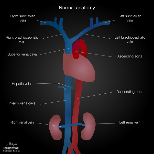
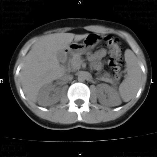
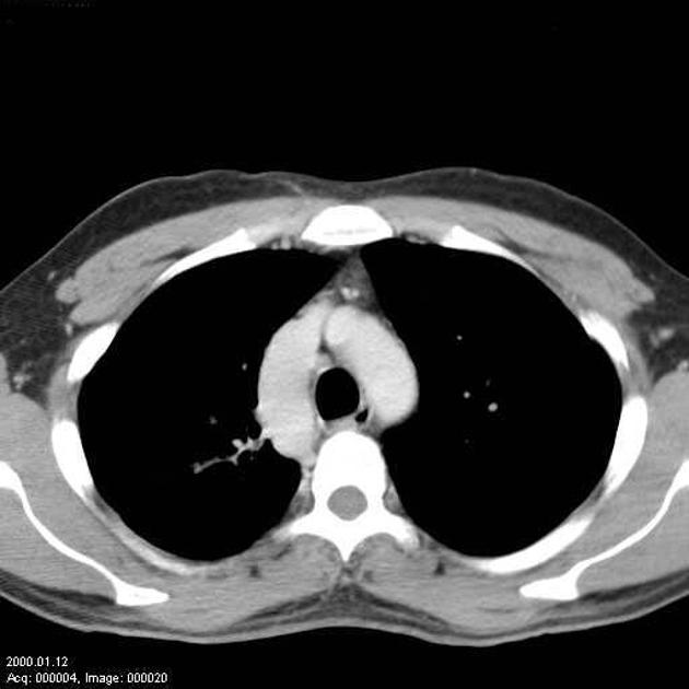
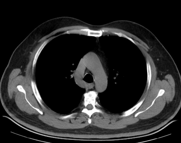
Caval variants, the variance of the anatomy of the venae cavae are common, due to the complex embryology of the venous system. Caval variants are important for a number of reasons:
to avoid confusion with venous pathology
to suggest the presence of frequently associated abnormalities
to plan vascular intervention/surgery
Types
-
superior vena cava (SVC)
-
inferior vena cava (IVC)
-
postrenal
persistent right posterior cardinal vein (retrocaval or circumcaval ureter)
persistent left supracardinal vein (transposition of IVC or left-sided IVC)
persistent bilateral supracardinal veins (duplication of IVC)
-
renal
-
prerenal
-
See also
References
- 1. Bass JE, Redwine MD, Kramer LA et-al. Spectrum of congenital anomalies of the inferior vena cava: cross-sectional imaging findings. Radiographics. 20 (3): 639-52. Radiographics (full text) - Pubmed citation
- 2. Kellman GM, Alpern MB, Sandler MA et-al. Computed tomography of vena caval anomalies with embryologic correlation. Radiographics. 1988;8 (3): 533-56. Radiographics (abstract) - Pubmed citation
- 3. Padhani AR, Hale HL. Mediastinal venous anomalies: potential pitfalls in cancer diagnosis. Br J Radiol. 1998;71 (847): 792-8. Br J Radiol (abstract) - Pubmed citation
- 4. Chuang VP, Mena CE, Hoskins PA. Congenital anomalies of the inferior vena cava. Review of embryogenesis and presentation of a simplified classification. Br J Radiol. 1974;47 (556): 206-13. Br J Radiol (citation) - Pubmed citation
- 5. Bass JE, Redwine MD, Kramer LA, Huynh PT, Harris JH. Spectrum of congenital anomalies of the inferior vena cava: cross-sectional imaging findings. Radiographics : a review publication of the Radiological Society of North America, Inc. 20 (3): 639-52. doi:10.1148/radiographics.20.3.g00ma09639 - Pubmed
Incoming Links
- Left sided inferior vena cava with azygos continuation
- Azygos continuation of inferior vena cava
- Left sided superior vena cava
- Left-sided superior vena cava
- Absent infrarenal inferior vena cava
- Posterior nutcracker phenomenon - retroaortic left renal vein draining into IVC
- Azygos continuation of the inferior vena cava
- Inferior vena cava duplication
- Caval variants (illustrations)
- Congenital absence of the IVC
- Left-sided inferior vena cava
- Persistent left superior vena cava
- Double inferior vena cava
- Duplication of the inferior vena cava
Related articles: Anatomy: Thoracic
- thoracic skeleton[+][+]
- thoracic cage
- thoracic spine
- articulations
- muscles of the thorax[+][+]
- diaphragm
- intercostal space
- intercostal muscles
- variant anatomy
- spaces of the thorax[+][+]
- thoracic viscera[+][+]
- lower respiratory tract
-
heart
- cardiac chambers
- heart valves
- cardiac fibrous skeleton
- innervation of the heart
- development of the heart
- cardiac wall
-
pericardium
- epicardium
- epicardial fat pad
- pericardial space
- oblique pericardial sinus
- transverse pericardial sinus
-
pericardial recesses
- aortic recesses
- pulmonic recesses
- postcaval recess
- pulmonary venous recesses
- pericardial ligaments
- myocardium
- endocardium
-
pericardium
- esophagus
- thymus
- breast
- arterial supply of the thorax[+][+]
-
thoracic aorta (development)
-
ascending aorta
-
aortic root
- aortic annulus
-
coronary arteries
- coronary arterial dominance
- myocardial segments
-
left main coronary artery (LMCA)
- ramus intermedius artery (RI)
-
circumflex artery (LCx)
- obtuse marginal branches (OM1, OM2, etc))
- Kugel's artery
-
left anterior descending artery (LAD)
- diagonal branches (D1, D2, etc)
- septal perforators (S1, S2, etc)
-
right coronary artery (RCA)
- conus artery
- sinoatrial nodal artery
- acute marginal branches (AM1, AM2, etc)
- inferior interventricular artery (PDA)
- posterior left ventricular artery (PLV)
- congenital anomalies
- sinotubular junction
-
aortic root
- aortic arch
- aortic isthmus
- descending aorta
-
ascending aorta
- pulmonary trunk
-
thoracic aorta (development)
- venous drainage of the thorax
-
superior vena cava (SVC)
- superior cavoatrial junction
- variant anatomy
- inferior vena cava (IVC)[+][+]
-
coronary veins[+][+]
-
cardiac veins which drain into the coronary sinus
- great cardiac vein
- middle cardiac vein
- small cardiac vein
- posterior vein of the left ventricle
- vein of Marshall (oblique vein of the left atrium)
- anterior cardiac veins
- venae cordis minimae (smallest cardiac veins or thebesian veins)
-
cardiac veins which drain into the coronary sinus
- pulmonary veins
- bronchial veins
- thoracoepigastric vein
-
superior vena cava (SVC)
- lymphatics of the thorax[+][+]
- innervation of the thorax[+][+]
Related articles: Anatomy: Abdominopelvic
- skeleton of the abdomen and pelvis
- muscles of the abdomen and pelvis
- spaces of the abdomen and pelvis
- anterior abdominal wall
- posterior abdominal wall
- abdominal cavity
- pelvic cavity
- perineum
- abdominal and pelvic viscera
- gastrointestinal tract
- spleen
- hepatobiliary system
-
endocrine system
-
adrenal gland
- adrenal vessels
- chromaffin cells
- variants
- pancreas
- organs of Zuckerkandl
-
adrenal gland
-
urinary system
-
kidney
- renal pelvis
- renal sinus
- avascular plane of Brodel
-
variants
- number
- fusion
- location
- shape
- ureter
- urinary bladder
- urethra
- embryology
-
kidney
- male reproductive system
-
female reproductive system
- vulva
- vagina
- uterus
- adnexa
- Fallopian tubes
- ovaries
- broad ligament (mnemonic)
- variant anatomy
- embryology
- blood supply of the abdomen and pelvis
- arteries
-
abdominal aorta
- inferior phrenic artery
- celiac artery
- superior mesenteric artery
- middle suprarenal artery
- renal artery (variant anatomy)
- gonadal artery (ovarian artery | testicular artery)
- inferior mesenteric artery
- lumbar arteries
- median sacral artery
-
common iliac artery
- external iliac artery
-
internal iliac artery (mnemonic)
- anterior division
- umbilical artery
- superior vesical artery
- obturator artery
- vaginal artery
- inferior vesical artery
- uterine artery
- middle rectal artery
-
internal pudendal artery
- inferior rectal artery
-
perineal artery
- posterior scrotal artery
- transverse perineal artery
- artery to the bulb
- deep artery of the penis/clitoris
- dorsal artery of the penis/clitoris
- inferior gluteal artery
- posterior division (mnemonic)
- variant anatomy
- anterior division
-
abdominal aorta
- portal venous system
- veins
- anastomoses
- arterioarterial anastomoses
- portal-systemic venous collateral pathways
- watershed areas
- arteries
- lymphatics
- innervation of the abdomen and pelvis
- thoracic splanchnic nerves
- lumbar plexus
-
sacral plexus
- lumbosacral trunk
- sciatic nerve
- superior gluteal nerve
- inferior gluteal nerve
- nerve to piriformis
- perforating cutaneous nerve
- posterior femoral cutaneous nerve
- parasympathetic pelvic splanchnic nerves
- pudendal nerve
- nerve to quadratus femoris and inferior gemellus muscles
- nerve to internal obturator and superior gemellus muscles
- autonomic ganglia and plexuses




 Unable to process the form. Check for errors and try again.