
Anterior mediastinum
Citation, DOI, disclosures and article data
Citation:
Jones J, Chieng R, Hacking C, et al. Anterior mediastinum. Reference article, Radiopaedia.org (Accessed on 20 Mar 2025) https://doi.org/10.53347/rID-14550
Permalink:
rID:
14550
Article created:
Disclosures:
At the time the article was created Jeremy Jones had no recorded disclosures.
View Jeremy Jones's current disclosures
Last revised:
Disclosures:
At the time the article was last revised Raymond Chieng had no financial relationships to ineligible companies to disclose.
View Raymond Chieng's current disclosures
Revisions:
10 times, by
7 contributors -
see full revision history and disclosures
Systems:
Sections:
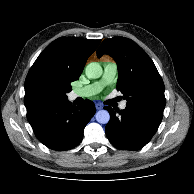
The anterior mediastinum is the portion of the mediastinum anterior to the pericardium and below the thoracic plane.
It forms the anterior part of the inferior mediastinum, and contains the thymus, lymph nodes, mammary vessels 3. It may contain the portions of a retrosternal thyroid.
Related pathology
The commonest pathology encountered within the anterior mediastinum is an anterior mediastinal mass. However, diffuse mediastinal disease may also occur.
References
- 1. Moore KL, Agur AMR, Dalley AF. Clinically oriented anatomy. LWW. ISBN:1451119453. Read it at Google Books - Find it at Amazon
- 2. Brett W. Carter, Marcelo F. Benveniste, Rachna Madan, Myrna C. Godoy, Patricia M. de Groot, Mylene T. Truong, Melissa L. Rosado-de-Christenson, Edith M. Marom. ITMIG Classification of Mediastinal Compartments and Multidisciplinary Approach to Mediastinal Masses. (2017) RadioGraphics. 37 (2): 413-436. doi:10.1148/rg.2017160095 - Pubmed
- 3. Stephanie Ryan, Michelle McNicholas, Stephen J. Eustace. Anatomy for Diagnostic Imaging. (2011) ISBN: 9780702029714 - Google Books
Incoming Links
Articles:
- Posterior mediastinum
- Anterior mediastinal mass in the exam
- Mediastinal seminoma
- Anterior mediastinal mass
- Parathyroid 4D CT
- Inferior mediastinum
- Mediastinal lymphoma
- Mediastinal teratoma
- Thymic epithelial tumours
- Manubrium
- Superior mediastinum
- Mediastinal embryonal carcinoma
- Middle mediastinum
- Mediastinum
- Testicular seminoma
- Mediastinal choriocarcinoma
- Paediatric mediastinal masses
- Mixed germ cell tumour of the mediastinum
Related articles: Anatomy: Thoracic
- thoracic skeleton[+][+]
- thoracic cage
- thoracic spine
- articulations
- muscles of the thorax[+][+]
- diaphragm
- intercostal space
- intercostal muscles
- variant anatomy
- spaces of the thorax
- thoracic viscera[+][+]
- lower respiratory tract
-
heart
- cardiac chambers
- heart valves
- cardiac fibrous skeleton
- innervation of the heart
- development of the heart
- cardiac wall
-
pericardium
- epicardium
- epicardial fat pad
- pericardial space
- oblique pericardial sinus
- transverse pericardial sinus
-
pericardial recesses
- aortic recesses
- pulmonic recesses
- postcaval recess
- pulmonary venous recesses
- pericardial ligaments
- myocardium
- endocardium
-
pericardium
- esophagus
- thymus
- breast
- arterial supply of the thorax[+][+]
-
thoracic aorta (development)
-
ascending aorta
-
aortic root
- aortic annulus
-
coronary arteries
- coronary arterial dominance
- myocardial segments
-
left main coronary artery (LMCA)
- ramus intermedius artery (RI)
-
circumflex artery (LCx)
- obtuse marginal branches (OM1, OM2, etc))
- Kugel's artery
-
left anterior descending artery (LAD)
- diagonal branches (D1, D2, etc)
- septal perforators (S1, S2, etc)
-
right coronary artery (RCA)
- conus artery
- sinoatrial nodal artery
- acute marginal branches (AM1, AM2, etc)
- inferior interventricular artery (PDA)
- posterior left ventricular artery (PLV)
- congenital anomalies
- sinotubular junction
-
aortic root
- aortic arch
- aortic isthmus
- descending aorta
-
ascending aorta
- pulmonary trunk
-
thoracic aorta (development)
- venous drainage of the thorax[+][+]
- superior vena cava (SVC)
- inferior vena cava (IVC)
-
coronary veins
-
cardiac veins which drain into the coronary sinus
- great cardiac vein
- middle cardiac vein
- small cardiac vein
- posterior vein of the left ventricle
- vein of Marshall (oblique vein of the left atrium)
- anterior cardiac veins
- venae cordis minimae (smallest cardiac veins or thebesian veins)
-
cardiac veins which drain into the coronary sinus
- pulmonary veins
- bronchial veins
- thoracoepigastric vein
- lymphatics of the thorax[+][+]
- innervation of the thorax[+][+]


 Unable to process the form. Check for errors and try again.