
Srb anomaly
Citation, DOI, disclosures and article data
Citation:
Bell D, Ibrahim D, Sharma R, Srb anomaly. Reference article, Radiopaedia.org (Accessed on 24 Mar 2025) https://doi.org/10.53347/rID-70445
Permalink:
rID:
70445
Article created:
Disclosures:
At the time the article was created Daniel J Bell had no recorded disclosures.
View Daniel J Bell's current disclosures
Last revised:
Disclosures:
At the time the article was last revised Dalia Ibrahim had no financial relationships to ineligible companies to disclose.
View Dalia Ibrahim's current disclosures
Revisions:
3 times, by
3 contributors -
see full revision history and disclosures
Systems:
Sections:
Synonyms:
- Srb's anomaly
- Srb anomalies
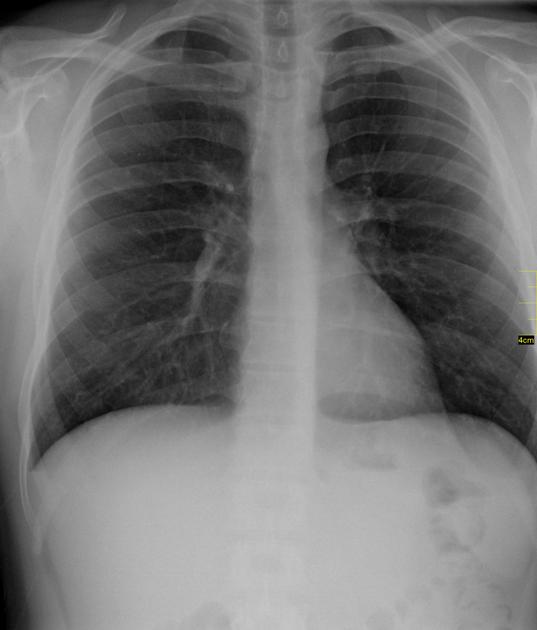
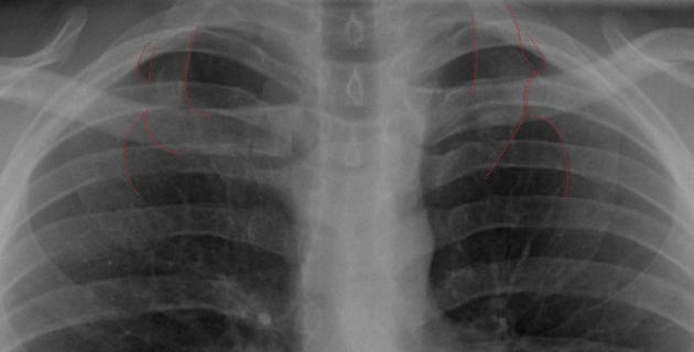
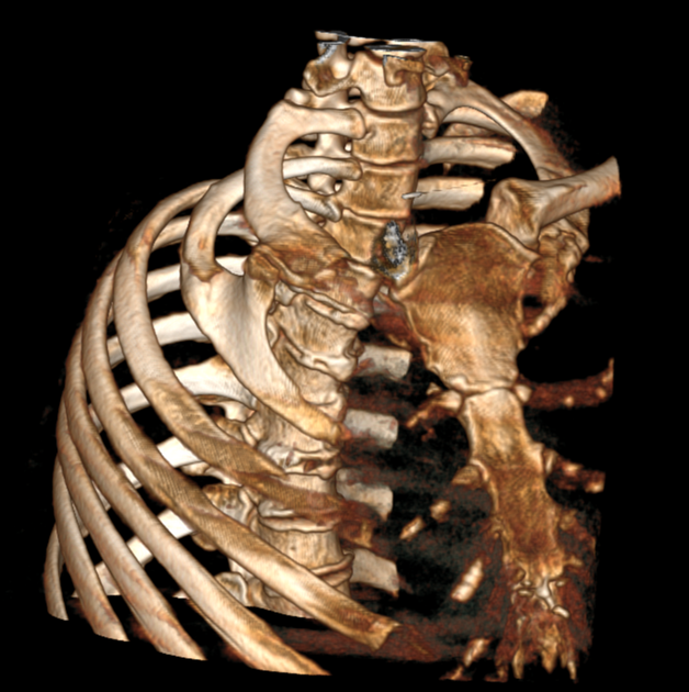
The Srb anomaly describes an anatomic variant of the ribs, in which there is partial to complete bony ankylosis of the first and second ribs.
References
- 1. Swischuk LE, Stansberry SD. Radiographic manifestations of anomalies of the chest wall. (1991) Radiologic clinics of North America. 29 (2): 271-8. Pubmed
- 2. Terry R. Yochum. Yochum and Rowe's Essentials of Skeletal Radiology. (2019) ISBN: 9780781739467
- 3. ROMANOWSKI B. [Srb's developmental anomaly complicated by neurological disorders]. (1961) Chirurgia narzadow ruchu i ortopedia polska. 26: 83-8. Pubmed
Incoming Links
Related articles: Anatomy: Thoracic
- thoracic skeleton
- thoracic cage
- thoracic spine
- articulations[+][+]
- muscles of the thorax[+][+]
- diaphragm
- intercostal space
- intercostal muscles
- variant anatomy
- spaces of the thorax[+][+]
- thoracic viscera[+][+]
- lower respiratory tract
-
heart
- cardiac chambers
- heart valves
- cardiac fibrous skeleton
- innervation of the heart
- development of the heart
- cardiac wall
-
pericardium
- epicardium
- epicardial fat pad
- pericardial space
- oblique pericardial sinus
- transverse pericardial sinus
-
pericardial recesses
- aortic recesses
- pulmonic recesses
- postcaval recess
- pulmonary venous recesses
- pericardial ligaments
- myocardium
- endocardium
-
pericardium
- esophagus
- thymus
- breast
- arterial supply of the thorax[+][+]
-
thoracic aorta (development)
-
ascending aorta
-
aortic root
- aortic annulus
-
coronary arteries
- coronary arterial dominance
- myocardial segments
-
left main coronary artery (LMCA)
- ramus intermedius artery (RI)
-
circumflex artery (LCx)
- obtuse marginal branches (OM1, OM2, etc))
- Kugel's artery
-
left anterior descending artery (LAD)
- diagonal branches (D1, D2, etc)
- septal perforators (S1, S2, etc)
-
right coronary artery (RCA)
- conus artery
- sinoatrial nodal artery
- acute marginal branches (AM1, AM2, etc)
- inferior interventricular artery (PDA)
- posterior left ventricular artery (PLV)
- congenital anomalies
- sinotubular junction
-
aortic root
- aortic arch
- aortic isthmus
- descending aorta
-
ascending aorta
- pulmonary trunk
-
thoracic aorta (development)
- venous drainage of the thorax[+][+]
- superior vena cava (SVC)
- inferior vena cava (IVC)
-
coronary veins
-
cardiac veins which drain into the coronary sinus
- great cardiac vein
- middle cardiac vein
- small cardiac vein
- posterior vein of the left ventricle
- vein of Marshall (oblique vein of the left atrium)
- anterior cardiac veins
- venae cordis minimae (smallest cardiac veins or thebesian veins)
-
cardiac veins which drain into the coronary sinus
- pulmonary veins
- bronchial veins
- thoracoepigastric vein
- lymphatics of the thorax[+][+]
- innervation of the thorax[+][+]


 Unable to process the form. Check for errors and try again.