
Inferior pulmonary ligament
Citation, DOI, disclosures and article data
At the time the article was created Henry Knipe had no recorded disclosures.
View Henry Knipe's current disclosuresAt the time the article was last revised Liz Silverstone had no financial relationships to ineligible companies to disclose.
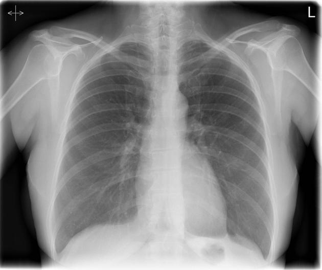
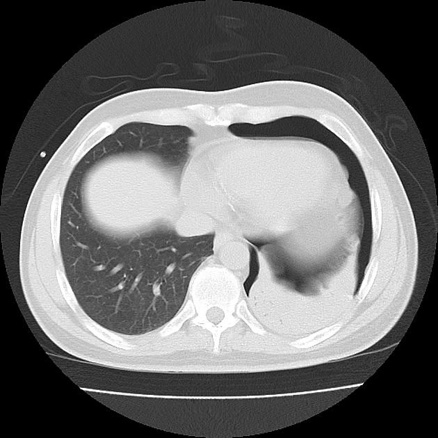
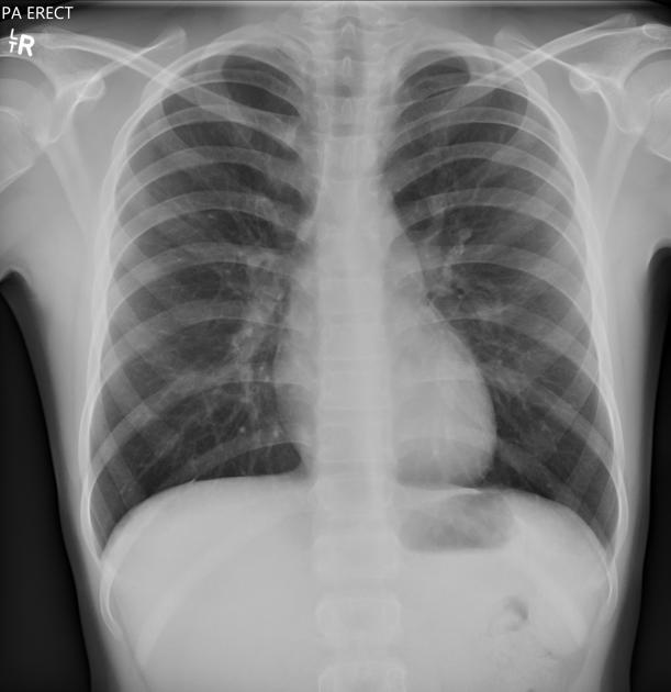
View Liz Silverstone's current disclosuresThe inferior pulmonary ligament (or pulmonary ligament) is not a true ligament; it is a normal and variable pleural fold which is sometimes seen on CT and may cause a triangular peak along the diaphragm on CXR.
On this page:
Gross anatomy
The inferior pulmonary ligament is formed by a double layer of visceral pleura with mediastinal pleura and connective tissue that extends from the root of the hilum towards the dome of the hemidiaphragm. It tethers the medial surface of the lower lobe to the mediastinum and allows hilar vessels to dilate when cardiac output is increased.
Contents
inferior pulmonary vein (at the apex) 1
Radiographic features
CT
thin, linear structure that inferior to the hilum on lung windows 3
most apparent at the level of the hemidiaphragm arising from the paraesophageal mediastinum 4
twice as commonly seen on the left than the right; evident in ~50% of patients 2, 3
ADVERTISEMENT: Supporters see fewer/no ads
Differential diagnosis
References
- 1. Brant WE, Helms C. Fundamentals of Diagnostic Radiology. Lippincott Williams & Wilkins. ISBN:1608319121. Read it at Google Books - Find it at Amazon
- 2. Rost RC, Proto AV. Inferior pulmonary ligament: computed tomographic appearance. Radiology. 1983;148 (2): 479-83. Pubmed citation
- 3. Cooper C, Moss AA, Buy JN et-al. CT appearance of the normal inferior pulmonary ligament. AJR Am J Roentgenol. 1983;141 (2): 237-40. doi:10.2214/ajr.141.2.237 - Pubmed citation
- 4. Andy Adam, Adrian K. Dixon, Cornelia Schaefer-Prokop et al. Grainger & Allison's Diagnostic Radiology, 2 Volume Set. (2020) ISBN: 9780702075247 - Google Books
Incoming Links
Related articles: Anatomy: Thoracic
- thoracic skeleton[+][+]
- thoracic cage
- thoracic spine
- articulations
- muscles of the thorax[+][+]
- diaphragm
- intercostal space
- intercostal muscles
- variant anatomy
- spaces of the thorax[+][+]
- thoracic viscera
-
lower respiratory tract
- tracheobronchial tree[+][+]
-
lungs
-
bronchopulmonary segmental anatomy (Boyden Classification) (mnemonic)[+][+]
- left lung
- right lung
- variant anatomy
- lung parenchyma[+][+]
-
hilum[+][+]
- pulmonary ligament
- pleura[+][+]
-
bronchopulmonary segmental anatomy (Boyden Classification) (mnemonic)[+][+]
-
heart[+][+]
- cardiac chambers
- heart valves
- cardiac fibrous skeleton
- innervation of the heart
- development of the heart
- cardiac wall
-
pericardium
- epicardium
- epicardial fat pad
- pericardial space
- oblique pericardial sinus
- transverse pericardial sinus
-
pericardial recesses
- aortic recesses
- pulmonic recesses
- postcaval recess
- pulmonary venous recesses
- pericardial ligaments
- myocardium
- endocardium
-
pericardium
- esophagus[+][+]
- thymus[+][+]
- breast[+][+]
-
lower respiratory tract
- arterial supply of the thorax[+][+]
-
thoracic aorta (development)
-
ascending aorta
-
aortic root
- aortic annulus
-
coronary arteries
- coronary arterial dominance
- myocardial segments
-
left main coronary artery (LMCA)
- ramus intermedius artery (RI)
-
circumflex artery (LCx)
- obtuse marginal branches (OM1, OM2, etc))
- Kugel's artery
-
left anterior descending artery (LAD)
- diagonal branches (D1, D2, etc)
- septal perforators (S1, S2, etc)
-
right coronary artery (RCA)
- conus artery
- sinoatrial nodal artery
- acute marginal branches (AM1, AM2, etc)
- inferior interventricular artery (PDA)
- posterior left ventricular artery (PLV)
- congenital anomalies
- sinotubular junction
-
aortic root
- aortic arch
- aortic isthmus
- descending aorta
-
ascending aorta
- pulmonary trunk
-
thoracic aorta (development)
- venous drainage of the thorax[+][+]
- superior vena cava (SVC)
- inferior vena cava (IVC)
-
coronary veins
-
cardiac veins which drain into the coronary sinus
- great cardiac vein
- middle cardiac vein
- small cardiac vein
- posterior vein of the left ventricle
- vein of Marshall (oblique vein of the left atrium)
- anterior cardiac veins
- venae cordis minimae (smallest cardiac veins or thebesian veins)
-
cardiac veins which drain into the coronary sinus
- pulmonary veins
- bronchial veins
- thoracoepigastric vein
- lymphatics of the thorax[+][+]
- innervation of the thorax[+][+]


 Unable to process the form. Check for errors and try again.