
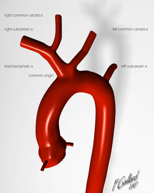
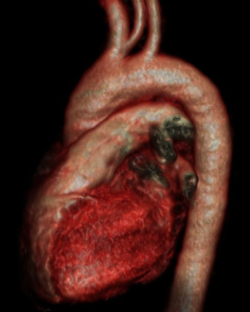
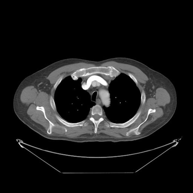
Bovine arch is the most common variant of the aortic arch and occurs when the brachiocephalic (innominate) artery shares a common origin with the left common carotid artery.
On this page:
Epidemiology
A bovine arch is present in ~15% (range 8-27%) of the population and is more common in individuals of African descent 1,7.
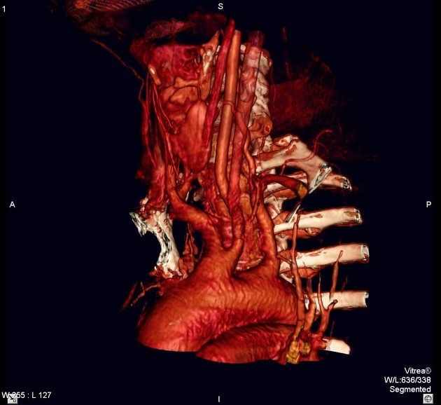
Gross anatomy
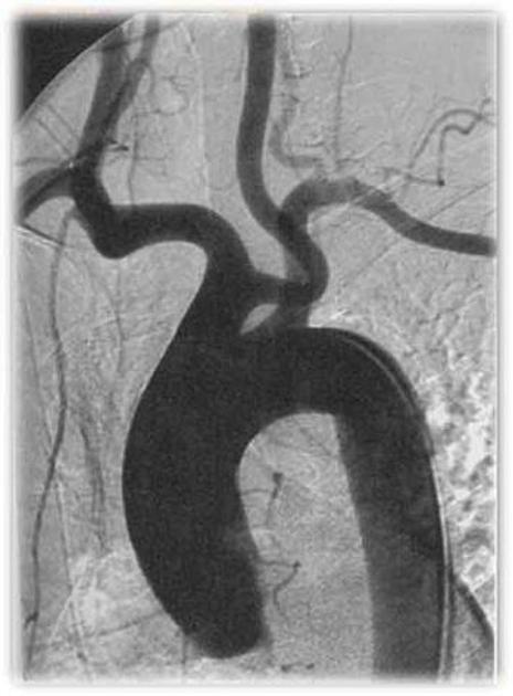
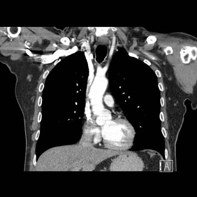
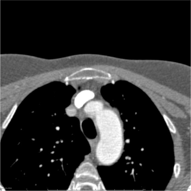
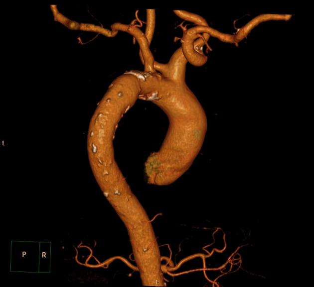
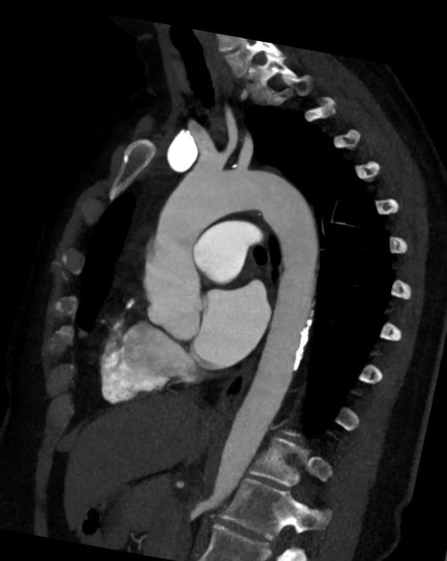
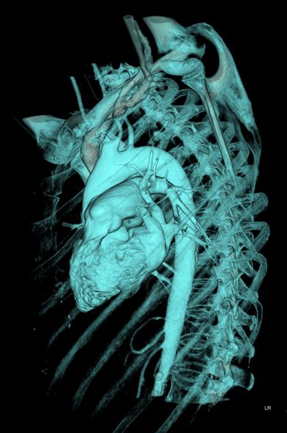
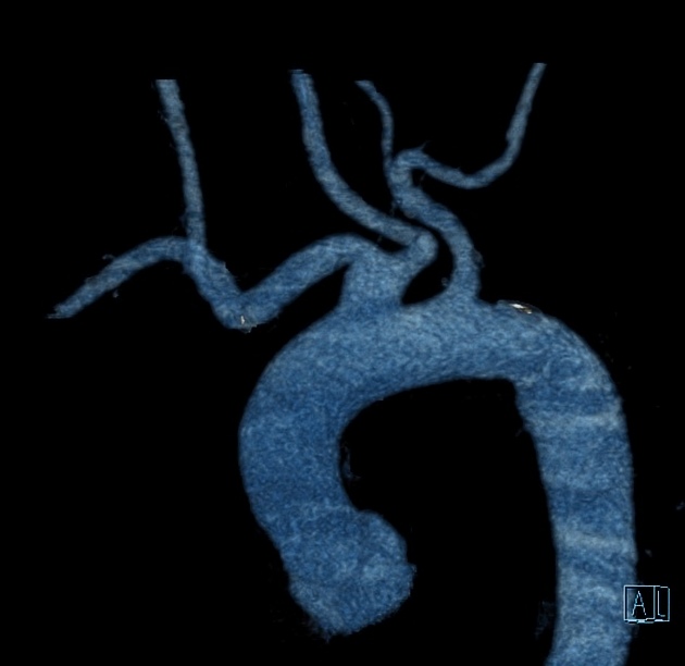
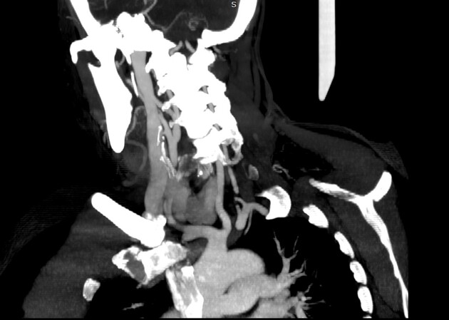
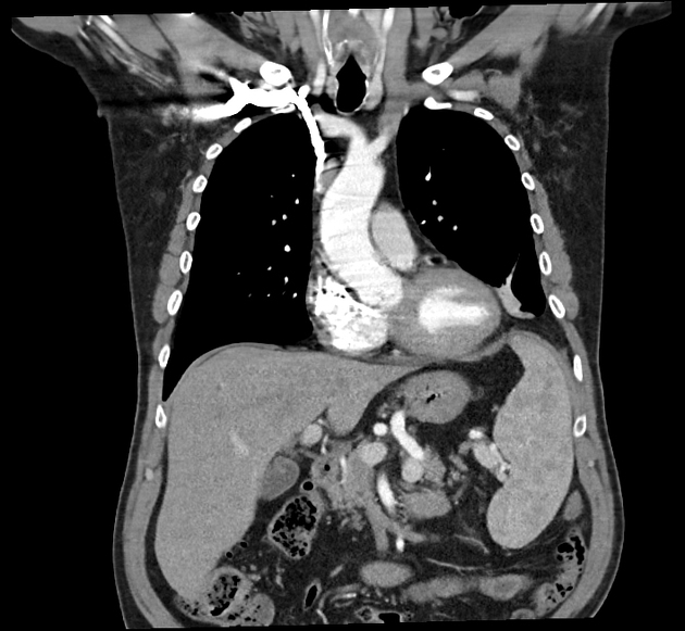
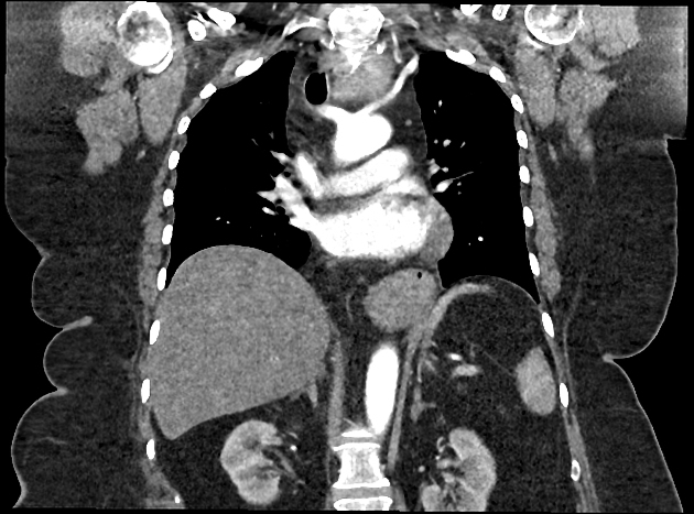
A bovine arch branching pattern is described as a common origin of the brachiocephalic artery and the left common carotid artery. The left subclavian artery arises independently from the more distal arch.
There is a continuous spectrum of aortic arch branching patterns between arterial origins that are clearly separate, to origins that are adjacent (or common), to those where there is clear common trunk before bifurcation.
A bovine arch with a lower origin of the left common carotid artery from the common trunk tends to result in a more "V-shaped" course of the distal vessels. A bovine arch with an origin of the left common carotid artery further distal on the common trunk typically results in the left common carotid artery having a more horizontal configuration that is close to 180° relative to the course of the brachiocephalic artery.
Clinical relevance
This common variant is asymptomatic most of the time. In rare cases of head and neck surgery, e.g. tracheostomy, it can be a risk factor for injury and cause complications 4. In combination with an aberrant right subclavian artery it can cause a dysphagia lusoria.
Variations in aortic branch anatomy, such as the bovine arch, are of significant importance to interventional neuroradiology. Historically, in order to minimize contrast use, an aortic DSA is typically not performed unless there are difficulties in engaging the head and neck vessels. One technique to engage the left common carotid artery is to slowly withdraw the catheter from the brachiocephalic artery. As it disengages from the brachiocephalic origin the catheter will typically slip into the left common carotid. When the vessel cannot be located this way, an arch aortogram can be performed to assist location of the vessels. An important clue to the presence of a bovine arch is the horizontal path of the proximal left common carotid artery. With the widespread use of cross sectional imaging the anatomy is now usually able to be identified prior to angiography.
Given the challenges in catheterization, it is perhaps unsurprising that the main clinical relevance of a bovine arch is an increase in complications reported during interventional procedures 6.
Bovine arch can be associated with an increased risk of embolic stroke, prevalent thoracic aortic arch abnormalities, thoracic aneurysm, and dilation, and a higher mortality rate in aortic dissection8.
History and etymology
The bovine arch is not the typical aortic arch pattern in bovines (cattle). Bovines have a common single brachiocephalic trunk which trifurcates into bilateral subclavian arteries and a single bicarotid trunk. This pattern is very rare in humans, but when it occurs, it is typically referred to as a true bovine arch.
There has been a debate about the origin of the name 1,5,6. Some authors, highlighting that the bovine arch pattern is not the normal aortic branching pattern found in bovines, concluded the arrangement described as "a bovine arch" is a misnomer. Their preferred terminology is "common origin of the brachiocephalic artery and left common carotid artery" 1.
The alternative view points out that there is no evidence that the description arose from the comparative anatomy with bovines 5,6. The pattern in bovines has been known since at least 1933. Bovine arch first appeared in the (human) literature in 1988. There are many ways that a name is linked to an animal, and the most common is that the appearance resembles that of an animal (e.g. the Scottie dog appearance, leaping dolphin sign or fishtail pancreas). In some cases the sign occurs because the human variant is the normal pattern in an animal, such as the pig bronchus (and, indeed, the true bovine arch).
In the case of the bovine arch, the 180° angle of the bifurcation resembles the 180° angle of the horns of a bovine. This is the likely origin of the term "bovine arch". The fact that the pattern does not occur in bovines does not render the term a misnomer. A butterfly vertebra, for example, would never be considered a misnomer even though butterflies do not have vertebrae 5.
For those interested in comparative anatomy, the bovine arch pattern is common in dogs, cats, rabbits and a number of primates 6.
Related variants
truncus bicaroticus: shared origin of the left and right common carotid arteries
true bovine arch: common origin of all four aortic arch vessels; the most common arrangement in bovines but very rare in humans


 Unable to process the form. Check for errors and try again.