
Aortic spindle
Last revised by Vincent Tatco
on 27 Jul 2022
Citation, DOI, disclosures and article data
Citation:
Knipe H, Tatco V, Weerakkody Y, Aortic spindle. Reference article, Radiopaedia.org (Accessed on 22 Mar 2025) https://doi.org/10.53347/rID-46017
Permalink:
rID:
46017
Article created:
Disclosures:
At the time the article was created Henry Knipe had no recorded disclosures.
View Henry Knipe's current disclosures
Last revised:
27 Jul 2022,
Vincent Tatco
Disclosures:
At the time the article was last revised Vincent Tatco had no recorded disclosures.
View Vincent Tatco's current disclosures
Revisions:
3 times, by
3 contributors -
see full revision history and disclosures
Systems:
Sections:
Tags:
Synonyms:
- Aortic spindles
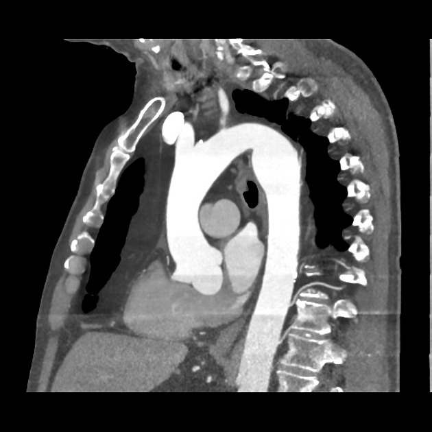
Aortic spindles are an anatomical variant of the proximal descending thoracic aorta. It occurs just distal to the aortic isthmus and has a circumferential smooth bulging appearance.
Differential diagnosis
- ductus diverticulum: not circumferential
- aortic pseudoaneurysm
- thoracic aortic aneurysm
References
- 1. Fisher RG, Sanchez-Torres M, Whigham CJ et-al. "Lumps" and "bumps" that mimic acute aortic and brachiocephalic vessel injury. Radiographics. 1997;17 (4): 825-34. doi:10.1148/radiographics.17.4.9225385 - Pubmed citation
- 2. Agarwal PP, Chughtai A, Matzinger FR et-al. Multidetector CT of thoracic aortic aneurysms. Radiographics. 2009;29 (2): 537-52. doi:10.1148/rg.292075080 - Pubmed citation
- 3. Rajiah P. CT and MRI in the Evaluation of Thoracic Aortic Diseases. Int J Vasc Med. 2013;2013: 797189. doi:10.1155/2013/797189 - Free text at pubmed - Pubmed citation
Incoming Links
Related articles: Anatomy: Thoracic
- thoracic skeleton[+][+]
- thoracic cage
- thoracic spine
- articulations
- muscles of the thorax[+][+]
- diaphragm
- intercostal space
- intercostal muscles
- variant anatomy
- spaces of the thorax[+][+]
- thoracic viscera[+][+]
- lower respiratory tract
-
heart
- cardiac chambers
- heart valves
- cardiac fibrous skeleton
- innervation of the heart
- development of the heart
- cardiac wall
-
pericardium
- epicardium
- epicardial fat pad
- pericardial space
- oblique pericardial sinus
- transverse pericardial sinus
-
pericardial recesses
- aortic recesses
- pulmonic recesses
- postcaval recess
- pulmonary venous recesses
- pericardial ligaments
- myocardium
- endocardium
-
pericardium
- esophagus
- thymus
- breast
- arterial supply of the thorax
-
thoracic aorta (development)
-
ascending aorta[+][+]
-
aortic root
- aortic annulus
-
coronary arteries
- coronary arterial dominance
- myocardial segments
-
left main coronary artery (LMCA)
- ramus intermedius artery (RI)
-
circumflex artery (LCx)
- obtuse marginal branches (OM1, OM2, etc))
- Kugel's artery
-
left anterior descending artery (LAD)
- diagonal branches (D1, D2, etc)
- septal perforators (S1, S2, etc)
-
right coronary artery (RCA)
- conus artery
- sinoatrial nodal artery
- acute marginal branches (AM1, AM2, etc)
- inferior interventricular artery (PDA)
- posterior left ventricular artery (PLV)
- congenital anomalies
- sinotubular junction
-
aortic root
- aortic arch[+][+]
-
aortic isthmus
- aortic spindle
- ductus arteriosum
- ligamentum arteriosum
- descending aorta[+][+]
-
ascending aorta[+][+]
- pulmonary trunk[+][+]
-
thoracic aorta (development)
- venous drainage of the thorax[+][+]
- superior vena cava (SVC)
- inferior vena cava (IVC)
-
coronary veins
-
cardiac veins which drain into the coronary sinus
- great cardiac vein
- middle cardiac vein
- small cardiac vein
- posterior vein of the left ventricle
- vein of Marshall (oblique vein of the left atrium)
- anterior cardiac veins
- venae cordis minimae (smallest cardiac veins or thebesian veins)
-
cardiac veins which drain into the coronary sinus
- pulmonary veins
- bronchial veins
- thoracoepigastric vein
- lymphatics of the thorax[+][+]
- innervation of the thorax[+][+]


 Unable to process the form. Check for errors and try again.