
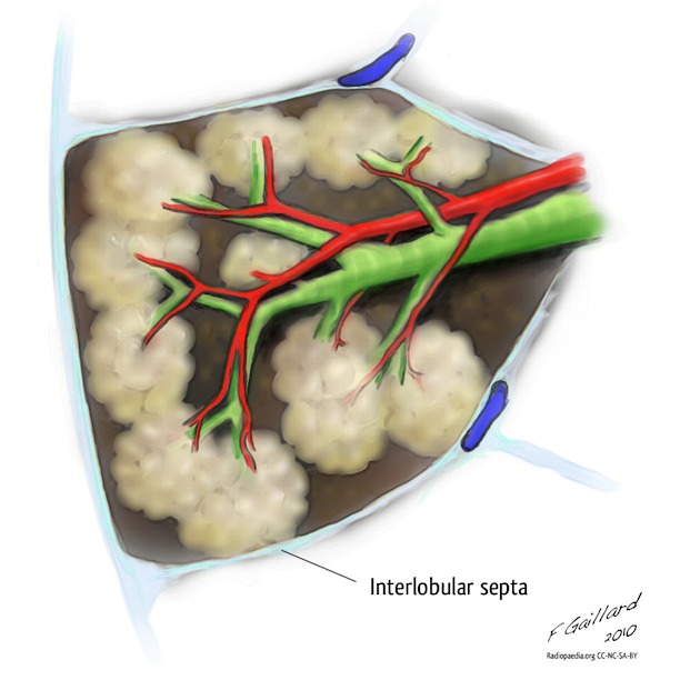
Interlobular septa
Citation, DOI, disclosures and article data
At the time the article was created Frank Gaillard had no recorded disclosures.
View Frank Gaillard's current disclosuresAt the time the article was last revised Daniel J Bell had no financial relationships to ineligible companies to disclose.
View Daniel J Bell's current disclosures- Interlobular interstitium
- Interlobular septum
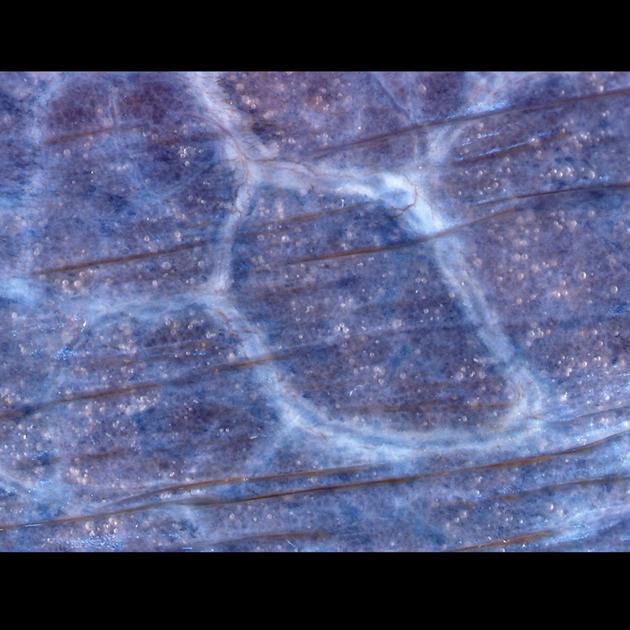
The interlobular septa (singular: interlobular septum) are located between the secondary pulmonary lobules and are continuous with both the subpleural interstitium (peripheral connective tissue) and the peribronchovascular interstitium (axial connective tissue) as well as the more delicate intralobular septa.
These septa are composed of connective tissues within which run the pulmonary veins and lymphatics which drain towards the pleura. A second set of lymphatics runs along with the arteries and drains centrally.
The interlobular septa are incomplete allowing for communication between adjacent secondary pulmonary lobules (canals of Lambert and pores of Kohn).
Related pathology
See also
References
- 1. Griffin CB, Primack SL. High-resolution CT: normal anatomy, techniques, and pitfalls. Radiol. Clin. North Am. 2001;39 (6): 1073-90, v. Pubmed citation
- 2. Verschakelen JA, de Wever W. Computed Tomography of the Lung: A Pattern Approach. Springer. ISBN:3540261877. Read it at Google Books - Find it at Amazon
Incoming Links
Related articles: Anatomy: Thoracic
- thoracic skeleton[+][+]
- thoracic cage
- thoracic spine
- articulations
- muscles of the thorax[+][+]
- diaphragm
- intercostal space
- intercostal muscles
- variant anatomy
- spaces of the thorax[+][+]
- thoracic viscera
-
lower respiratory tract
- tracheobronchial tree[+][+]
-
lungs
-
bronchopulmonary segmental anatomy (Boyden Classification) (mnemonic)[+][+]
- left lung
- right lung
- variant anatomy
- lung parenchyma
- hilum[+][+]
- pleura[+][+]
-
bronchopulmonary segmental anatomy (Boyden Classification) (mnemonic)[+][+]
-
heart[+][+]
- cardiac chambers
- heart valves
- cardiac fibrous skeleton
- innervation of the heart
- development of the heart
- cardiac wall
-
pericardium
- epicardium
- epicardial fat pad
- pericardial space
- oblique pericardial sinus
- transverse pericardial sinus
-
pericardial recesses
- aortic recesses
- pulmonic recesses
- postcaval recess
- pulmonary venous recesses
- pericardial ligaments
- myocardium
- endocardium
-
pericardium
- esophagus[+][+]
- thymus[+][+]
- breast[+][+]
-
lower respiratory tract
- arterial supply of the thorax[+][+]
-
thoracic aorta (development)
-
ascending aorta
-
aortic root
- aortic annulus
-
coronary arteries
- coronary arterial dominance
- myocardial segments
-
left main coronary artery (LMCA)
- ramus intermedius artery (RI)
-
circumflex artery (LCx)
- obtuse marginal branches (OM1, OM2, etc))
- Kugel's artery
-
left anterior descending artery (LAD)
- diagonal branches (D1, D2, etc)
- septal perforators (S1, S2, etc)
-
right coronary artery (RCA)
- conus artery
- sinoatrial nodal artery
- acute marginal branches (AM1, AM2, etc)
- inferior interventricular artery (PDA)
- posterior left ventricular artery (PLV)
- congenital anomalies
- sinotubular junction
-
aortic root
- aortic arch
- aortic isthmus
- descending aorta
-
ascending aorta
- pulmonary trunk
-
thoracic aorta (development)
- venous drainage of the thorax[+][+]
- superior vena cava (SVC)
- inferior vena cava (IVC)
-
coronary veins
-
cardiac veins which drain into the coronary sinus
- great cardiac vein
- middle cardiac vein
- small cardiac vein
- posterior vein of the left ventricle
- vein of Marshall (oblique vein of the left atrium)
- anterior cardiac veins
- venae cordis minimae (smallest cardiac veins or thebesian veins)
-
cardiac veins which drain into the coronary sinus
- pulmonary veins
- bronchial veins
- thoracoepigastric vein
- lymphatics of the thorax[+][+]
- innervation of the thorax[+][+]


 Unable to process the form. Check for errors and try again.