
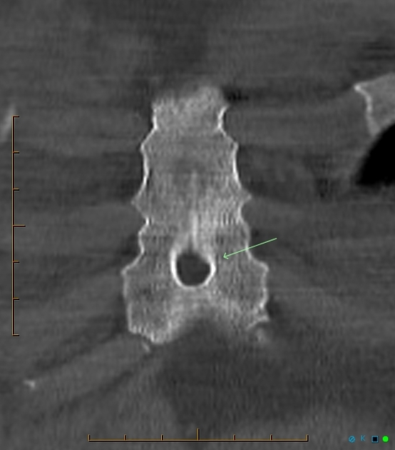
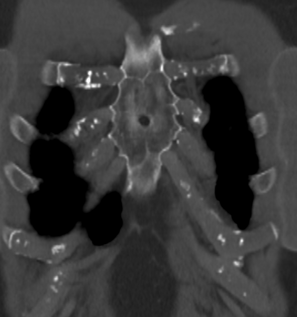
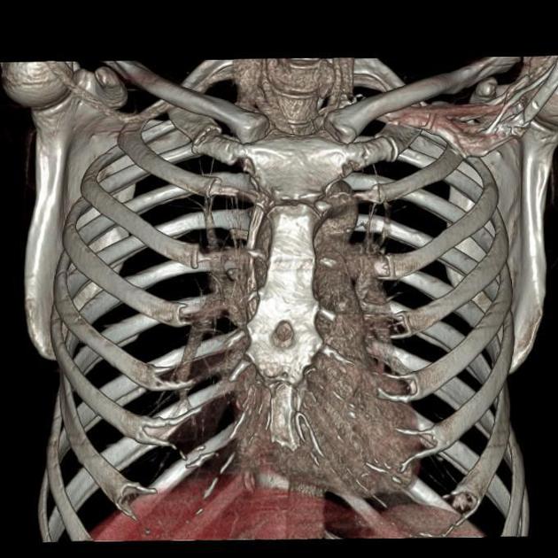
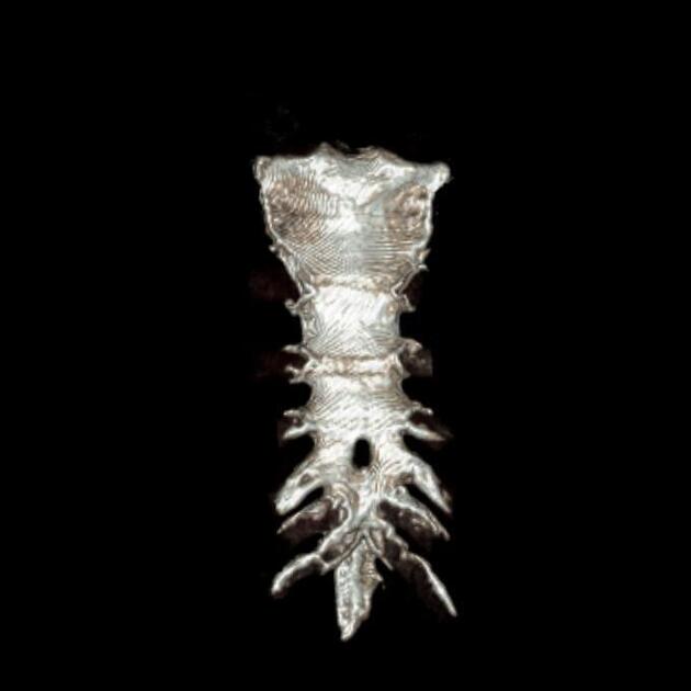
Sternal foramen
Citation, DOI, disclosures and article data
At the time the article was created Henry Knipe had no recorded disclosures.
View Henry Knipe's current disclosuresAt the time the article was last revised Liz Silverstone had no financial relationships to ineligible companies to disclose.
View Liz Silverstone's current disclosures- Perforated sternum
- Sternal foramina
Sternal foramen (or perforated sternum) is a developmental variant of the sternum and results from incomplete fusion of the sternal ossification centers. Xiphoid foramen can also occur, but is of lesser clinical significance 4.
On this page:
Epidemiology
They are common, occurring in approximately 5% of the population (range 4.3-6.7%).
Diagnosis
Sternal foramina are most commonly asymptomatic incidental findings on CT, typically found in the inferior portion of the sternal body.
Pathology
Sternal foraminae result from incomplete fusion of multiple right and left primary ossification centers in a single sternebra and are most frequent from the 2nd to 4th sternebrae. The size can vary from 2-16 mm and the defect is typically impalpable.
Differential diagnosis
Small foramina could be mistaken for sternal fractures.
Practical points
Blind interventions such as marrow biopsy could injure the pericardium, right ventricle, aorta or lung 5.
References
- 1. Gossner J. Relationship of sternal foramina to vital structures of the chest: a computed tomographic study. Anat Res Int. 2013;2013: 780193. doi:10.1155/2013/780193 - Free text at pubmed - Pubmed citation
- 2. Tarladacalisir T, Karamustafaoglu YA. A rare entity: sternal foramen. Eur J Cardiothorac Surg. 2013;44 (2): 384. doi:10.1093/ejcts/ezt091 - Pubmed citation
- 3. AKTAN, Zühre Aslı. "Anatomic and HRCT demonstration of midline sternal foramina." Turkish Journal of Medical Sciences 28.5 (1998): 511-514.
- 4. Chary Duraikannu, Olma V Noronha, Pushparajan Sundarrajan. MDCT evaluation of sternal variations: Pictorial essay. (2016) Indian Journal of Radiology and Imaging. 26 (2): 185. doi:10.4103/0971-3026.184407 - Pubmed
- 5. Choi P, Iwanaga J, Tubbs R. A Comprehensive Review of the Sternal Foramina and Its Clinical Significance. Cureus. 2017;9(12):e1929. doi:10.7759/cureus.1929 - Pubmed
Incoming Links
Related articles: Anatomy: Thoracic
- thoracic skeleton
- thoracic cage
- thoracic spine
- articulations[+][+]
- muscles of the thorax[+][+]
- diaphragm
- intercostal space
- intercostal muscles
- variant anatomy
- spaces of the thorax[+][+]
- thoracic viscera[+][+]
- lower respiratory tract
-
heart
- cardiac chambers
- heart valves
- cardiac fibrous skeleton
- innervation of the heart
- development of the heart
- cardiac wall
-
pericardium
- epicardium
- epicardial fat pad
- pericardial space
- oblique pericardial sinus
- transverse pericardial sinus
-
pericardial recesses
- aortic recesses
- pulmonic recesses
- postcaval recess
- pulmonary venous recesses
- pericardial ligaments
- myocardium
- endocardium
-
pericardium
- esophagus
- thymus
- breast
- arterial supply of the thorax[+][+]
-
thoracic aorta (development)
-
ascending aorta
-
aortic root
- aortic annulus
-
coronary arteries
- coronary arterial dominance
- myocardial segments
-
left main coronary artery (LMCA)
- ramus intermedius artery (RI)
-
circumflex artery (LCx)
- obtuse marginal branches (OM1, OM2, etc))
- Kugel's artery
-
left anterior descending artery (LAD)
- diagonal branches (D1, D2, etc)
- septal perforators (S1, S2, etc)
-
right coronary artery (RCA)
- conus artery
- sinoatrial nodal artery
- acute marginal branches (AM1, AM2, etc)
- inferior interventricular artery (PDA)
- posterior left ventricular artery (PLV)
- congenital anomalies
- sinotubular junction
-
aortic root
- aortic arch
- aortic isthmus
- descending aorta
-
ascending aorta
- pulmonary trunk
-
thoracic aorta (development)
- venous drainage of the thorax[+][+]
- superior vena cava (SVC)
- inferior vena cava (IVC)
-
coronary veins
-
cardiac veins which drain into the coronary sinus
- great cardiac vein
- middle cardiac vein
- small cardiac vein
- posterior vein of the left ventricle
- vein of Marshall (oblique vein of the left atrium)
- anterior cardiac veins
- venae cordis minimae (smallest cardiac veins or thebesian veins)
-
cardiac veins which drain into the coronary sinus
- pulmonary veins
- bronchial veins
- thoracoepigastric vein
- lymphatics of the thorax[+][+]
- innervation of the thorax[+][+]


 Unable to process the form. Check for errors and try again.