
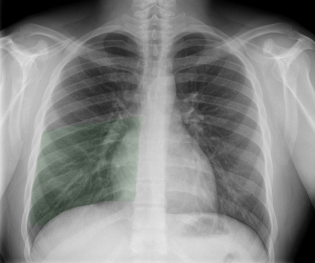
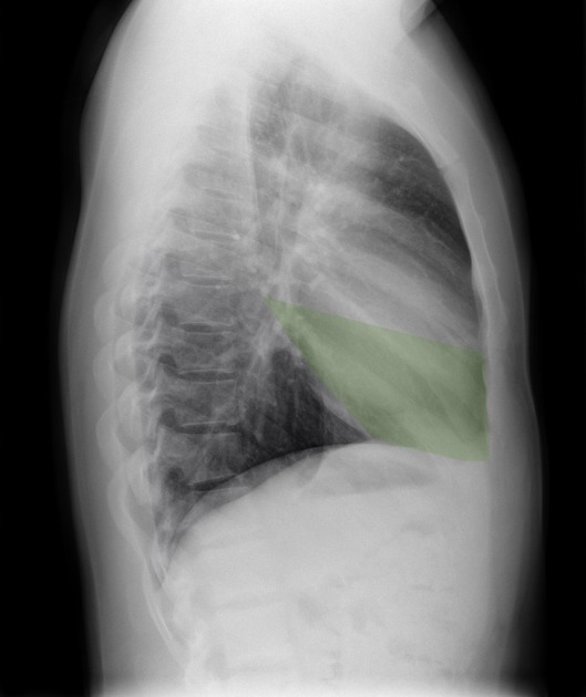
The right middle lobe (RML) or simply the middle lobe is one of three lobes in the right lung. It is separated from the right upper lobe above by the horizontal fissure and the right lower lobe below by the right oblique fissure and is subdivided into two bronchopulmonary segments.
On this page:
Gross anatomy
Location and structure
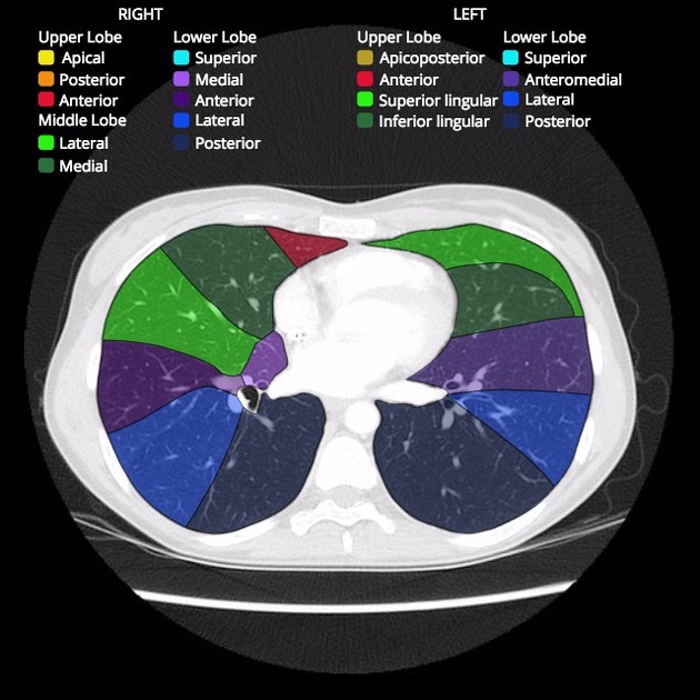
The right middle lobe lies in the lower anterior aspect of the right hemithorax and contains two bronchopulmonary segments:
lateral segment (segment 4)
medial segment (segment 5)
This is easy to remember as the middle lobe has medial and lateral segments.
Like all the pulmonary lobes, it is lined by visceral pleura which reflects at the pulmonary hilum where it is continuous with the parietal pleura. The right middle lobe bronchus arises from the lateral wall of the bronchus intermedius to traverse the right hilum into the right middle lobe.
The right middle lobe is separated from the right lower lobe below and posteriorly by the oblique fissure and from the right upper lobe above by the horizontal fissure.
Arterial supply
Like all the lobes of the lung, the right middle lobe has dual arterial supply:
deoxygenated blood from the right upper lobar pulmonary artery
oxygenated blood from branches of the right bronchial arteries
Venous drainage
Venous drainage of newly oxygenated blood is via the right inferior pulmonary vein into the left atrium.
Right bronchial veins drain into the azygos vein.
Lymphatic drainage
The superficial subpleural lymphatic plexus drains the lung parenchyma and visceral pleura to the bronchopulmonary (hilar) lymph nodes in the hilum.
The deep bronchopulmonary lymphatic plexus (in the bronchial submucosa and peribronchial interstitium) drains the root of the lung to hilar lymph nodes in the hilum.
The hilar lymph nodes then drain to the tracheobronchial lymph nodes.
Innervation
Innervation is derived from the pulmonary plexus:
parasympathetic fibers from the vagus nerve (CN X)
sympathetic fibers from the paravertebral sympathetic trunks


 Unable to process the form. Check for errors and try again.