
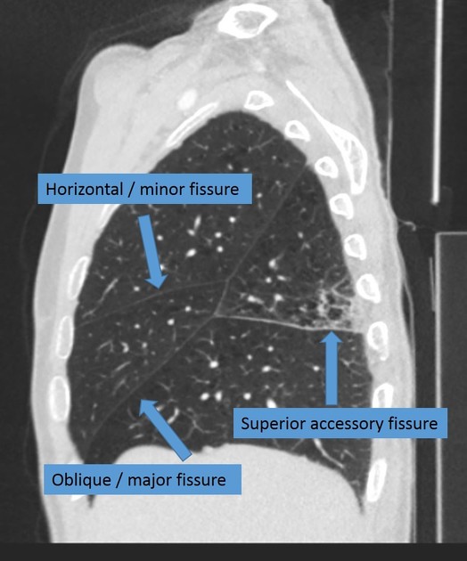
Superior accessory fissure
Citation, DOI, disclosures and article data
At the time the article was created Eric F Greif had no recorded disclosures.
View Eric F Greif's current disclosuresAt the time the article was last revised Raymond Chieng had no financial relationships to ineligible companies to disclose.
View Raymond Chieng's current disclosures- Superior accessory fissures
The superior accessory fissure is present in around 5% of individuals examined with CT 4 and PA chest X-rays 5.
On this page:
Gross anatomy
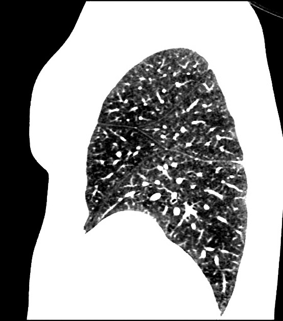
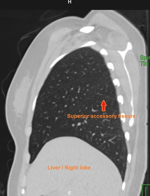
The superior accessory fissure of the right lower lobe is located in the same plane and posterior to the right transverse (minor) fissure. It separates the right lower lobe into superior (apical) and basal segments 5. This superior segment is called the posterior or dorsal lobe.
The fissure varies in length from complete to incomplete fissure 1,2. The incidence is greater with right lung involvement and less common than an inferior accessory fissure 1.
Radiographic features
Plain radiograph
Frontal chest radiographs demonstrate a thin horizontal linear opacity projecting below, parallel, and medial to the right minor fissure. The lateral radiograph demonstrates a thin line posterior to the right minor fissure, usually in the same plane and in a horizontal or oblique orientation. It may look like a continuation of the right minor fissure posteriorly, which separates the right lower lobe into superior and inferior segments 1,2.
Differential diagnosis
Consider:
pleural-parenchymal scarring
References
- 1. Godwin JD, Tarver RD. Accessory fissures of the lung. AJR Am J Roentgenol. 1985;144 (1): 39-47. doi:10.2214/ajr.144.1.39 - Pubmed citation
- 2. Goodman LR. Felson's Principles of Chest Roentgenology Text with CD-ROM. Saunders. ISBN:1416029230. Read it at Google Books - Find it at Amazon
- 3. Davis SD, Yu LS, Hentel KD. Obliquely oriented superior accessory fissure of the lower lobe of the lung: CT evaluation of the normal appearance and effect on the distribution of parenchymal and pleural opacities. Radiology. 2000;216 (1): 97-106. doi:10.1148/radiology.216.1.r00jl4797 - Pubmed citation
- 4. Yildiz A, Gölpinar F, Calikoğlu M, Duce MN, Ozer C, Apaydin FD. HRCT evaluation of the accessory fissures of the lung. European journal of radiology. 49 (3): 245-9. doi:10.1016/S0720-048X(03)00137-2 - Pubmed
- 5. Stephanie Ryan, Michelle McNicholas, Stephen J. Eustace. Anatomy for Diagnostic Imaging. (2011) Page 126. ISBN: 9780702029714 - Google Books
Incoming Links
Related articles: Anatomy: Thoracic
- thoracic skeleton[+][+]
- thoracic cage
- thoracic spine
- articulations
- muscles of the thorax[+][+]
- diaphragm
- intercostal space
- intercostal muscles
- variant anatomy
- spaces of the thorax[+][+]
- thoracic viscera
-
lower respiratory tract
- tracheobronchial tree[+][+]
-
lungs
-
bronchopulmonary segmental anatomy (Boyden Classification) (mnemonic)[+][+]
- left lung
- right lung
- variant anatomy
- lung parenchyma[+][+]
- hilum[+][+]
- pleura
-
bronchopulmonary segmental anatomy (Boyden Classification) (mnemonic)[+][+]
-
heart[+][+]
- cardiac chambers
- heart valves
- cardiac fibrous skeleton
- innervation of the heart
- development of the heart
- cardiac wall
-
pericardium
- epicardium
- epicardial fat pad
- pericardial space
- oblique pericardial sinus
- transverse pericardial sinus
-
pericardial recesses
- aortic recesses
- pulmonic recesses
- postcaval recess
- pulmonary venous recesses
- pericardial ligaments
- myocardium
- endocardium
-
pericardium
- esophagus[+][+]
- thymus[+][+]
- breast[+][+]
-
lower respiratory tract
- arterial supply of the thorax[+][+]
-
thoracic aorta (development)
-
ascending aorta
-
aortic root
- aortic annulus
-
coronary arteries
- coronary arterial dominance
- myocardial segments
-
left main coronary artery (LMCA)
- ramus intermedius artery (RI)
-
circumflex artery (LCx)
- obtuse marginal branches (OM1, OM2, etc))
- Kugel's artery
-
left anterior descending artery (LAD)
- diagonal branches (D1, D2, etc)
- septal perforators (S1, S2, etc)
-
right coronary artery (RCA)
- conus artery
- sinoatrial nodal artery
- acute marginal branches (AM1, AM2, etc)
- inferior interventricular artery (PDA)
- posterior left ventricular artery (PLV)
- congenital anomalies
- sinotubular junction
-
aortic root
- aortic arch
- aortic isthmus
- descending aorta
-
ascending aorta
- pulmonary trunk
-
thoracic aorta (development)
- venous drainage of the thorax[+][+]
- superior vena cava (SVC)
- inferior vena cava (IVC)
-
coronary veins
-
cardiac veins which drain into the coronary sinus
- great cardiac vein
- middle cardiac vein
- small cardiac vein
- posterior vein of the left ventricle
- vein of Marshall (oblique vein of the left atrium)
- anterior cardiac veins
- venae cordis minimae (smallest cardiac veins or thebesian veins)
-
cardiac veins which drain into the coronary sinus
- pulmonary veins
- bronchial veins
- thoracoepigastric vein
- lymphatics of the thorax[+][+]
- innervation of the thorax[+][+]
Related articles: Chest
- imaging techniques
-
chest radiograph
- radiography[+][+]
-
approach
- ABCDE
- ABCDEFGHI
- congenital heart disease
- medical devices in the thorax
- common lines and tubes[+][+]
- nasogastric tubes
- endotracheal tubes
- central venous catheters
- esophageal temperature probe
- tracheostomy tube
- pleural catheters
- cardiac conduction devices
- prosthetic heart valve
- review areas
-
airspace opacification[+][+]
- differential diagnoses of airspace opacification
- lobar consolidation
-
atelectasis
- mechanism-based
- morphology-based
- lobar lung collapse
- chest x-ray in the exam setting[+][+]
- cardiomediastinal contour[+][+]
- chest radiograph zones[+][+]
- tracheal air column[+][+]
-
fissures
- horizontal
- oblique
-
accessory fissures
- azygos fissure
- superior accessory fissure
- inferior accessory fissure
- left horizontal fissure
- vertical fissure line
- normal chest x-ray appearance of the diaphragm[+][+]
- nipple shadow[+][+]
-
lines and stripes[+][+]
- anterior junction line
- posterior junction line
- right paratracheal stripe
- left paratracheal stripe
- posterior tracheal stripe/tracheo-esophageal stripe
- posterior wall of bronchus intermedius
- right paraspinal line
- left paraspinal line
- aortic-pulmonary stripe
- aortopulmonary window
- azygo-esophageal recess
- spaces[+][+]
- signs[+][+]
- air bronchogram
- big rib sign
- Chang sign
- Chen sign
- coin lesion
- continuous diaphragm sign
- dense hilum sign
- double contour sign
- egg-on-a-string sign
- extrapleural sign
- finger in glove sign
- flat waist sign
- Fleischner sign
- ginkgo leaf sign
- Golden S sign
- Hampton hump
- haystack sign
- hilum convergence sign
- hilum overlay sign
- Hoffman-Rigler sign
- holly leaf sign
- incomplete border sign
- juxtaphrenic peak sign
- Kirklin sign
- medial stripe sign
- melting ice cube sign
- more black sign
- Naclerio V sign
- Palla sign
- pericardial fat tag sign
- Shmoo sign
- silhouette sign
- snowman sign
- spinnaker sign
- steeple sign
- straight left heart border sign
- third mogul sign
- tram-track sign
- walking man sign
- water bottle sign
- wave sign
- Westermark sign
- HRCT[+][+]
-
chest radiograph
- airways[+][+]
- bronchitis
- small airways disease
-
bronchiectasis
- broncho-arterial ratio
- related conditions
- differentials by distribution
- narrowing
-
tracheal stenosis
- diffuse tracheal narrowing (differential)
-
bronchial stenosis
- diffuse airway narrowing (differential)
-
tracheal stenosis
- diverticula
- pulmonary edema[+][+]
-
interstitial lung disease (ILD)[+][+]
- Anti-Jo-1 antibody-positive interstitial lung disease
- drug-induced interstitial lung disease
-
hypersensitivity pneumonitis
- acute hypersensitivity pneumonitis
- subacute hypersensitivity pneumonitis
- chronic hypersensitivity pneumonitis
- etiology
- bird fancier's lung: pigeon fancier's lung
- farmer's lung
- cheese workers' lung
- bagassosis
- mushroom worker’s lung
- malt worker’s lung
- maple bark disease
- hot tub lung
- wine maker’s lung
- woodsman’s disease
- thatched roof lung
- tobacco grower’s lung
- potato riddler’s lung
- summer-type pneumonitis
- dry rot lung
- machine operator’s lung
- humidifier lung
- shower curtain disease
- furrier’s lung
- miller’s lung
- lycoperdonosis
- saxophone lung
-
idiopathic interstitial pneumonia (mnemonic)
- acute interstitial pneumonia (AIP)
- cryptogenic organizing pneumonia (COP)
- desquamative interstitial pneumonia (DIP)
- non-specific interstitial pneumonia (NSIP)
- idiopathic pleuroparenchymal fibroelastosis
- lymphoid interstitial pneumonia (LIP)
- respiratory bronchiolitis–associated interstitial lung disease (RB-ILD)
- usual interstitial pneumonia / idiopathic pulmonary fibrosis (UIP/IPF)
-
pneumoconioses
- fibrotic
- non-fibrotic
-
lung cancer[+][+]
-
non-small-cell lung cancer
-
adenocarcinoma
- pre-invasive tumors
- minimally invasive tumors
- invasive tumors
- variants of invasive carcinoma
- described imaging features
- adenosquamous carcinoma
- large cell carcinoma
- primary sarcomatoid carcinoma of the lung
- squamous cell carcinoma
- salivary gland-type tumors
-
adenocarcinoma
- pulmonary neuroendocrine tumors
- preinvasive lesions
-
lung cancer invasion patterns
- tumor spread through air spaces (STAS)
- presence of non-lepidic patterns such as acinar, papillary, solid, or micropapillary
- myofibroblastic stroma associated with invasive tumor cells
- pleural invasion
- vascular invasion
- tumors by location
- benign neoplasms
- pulmonary metastases
- lung cancer screening
- lung cancer staging
-
non-small-cell lung cancer


 Unable to process the form. Check for errors and try again.