
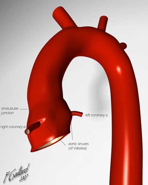
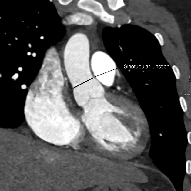
Sinotubular junction
Citation, DOI, disclosures and article data
Citation:
Gaillard F, Hacking C, Weerakkody Y, et al. Sinotubular junction. Reference article, Radiopaedia.org (Accessed on 08 Mar 2025) https://doi.org/10.53347/rID-2050
Permalink:
rID:
2050
Article created:
Disclosures:
At the time the article was created Frank Gaillard had no recorded disclosures.
View Frank Gaillard's current disclosures
Last revised:
Disclosures:
At the time the article was last revised Craig Hacking had the following disclosures:
- Philips Australia, Paid speaker at Philips Spectral CT events (ongoing)
These were assessed during peer review and were determined to not be relevant to the changes that were made.
View Craig Hacking's current disclosures
Revisions:
5 times, by
3 contributors -
see full revision history and disclosures
Systems:
Sections:
Synonyms:
- Sino-tubular junction
- STJ
The sinotubular junction (STJ) is the region of the ascending aorta between the aortic sinuses (of Valsalva) and where the normal tubular configuration of the aorta is attained. It marks the junction of the aortic root and ascending aorta.
References
- 1. Sinnatamby CS. Last's Anatomy, Regional And Applied. Churchill Livingstone. (2006) ISBN:0443100330. Read it at Google Books - Find it at Amazon
- 2. Clemente CD. Anatomy. Lippincott Williams & Wilkins. (2011) ISBN:1582558892. Read it at Google Books - Find it at Amazon
- 3. Bennett CJ, Maleszewski JJ, Araoz PA. CT and MR imaging of the aortic valve: radiologic-pathologic correlation. Radiographics. 2012;32 (5): 1399-420. doi:10.1148/rg.325115727 - Pubmed citation
- 4. Quint LE, Liu PS, Booher AM et-al. Proximal thoracic aortic diameter measurements at CT: repeatability and reproducibility according to measurement method. Int J Cardiovasc Imaging. 2013;29 (2): 479-88. doi:10.1007/s10554-012-0102-9 - Free text at pubmed - Pubmed citation
- 5. Ko JP, Goldstein JM, Latson LA, Azour L, Gozansky EK, Moore W, Patel S, Hutchinson B. Chest CT Angiography for Acute Aortic Pathologic Conditions: Pearls and Pitfalls. (2021) Radiographics : a review publication of the Radiological Society of North America, Inc. 41 (2): 399-424. doi:10.1148/rg.2021200055 - Pubmed
- 6.Charitos EI, Sievers HH. Anatomy of the aortic root: implications for valve-sparing surgery. (2013) Annals of cardiothoracic surgery. 2 (1): 53-6. doi:10.3978/j.issn.2225-319X.2012.11.18 - Pubmed
Incoming Links
Articles:
- Aortic root
- Transcatheter aortic valve implantation (TAVI)
- Tulip bulb sign
- Ascending aortic aneurysm
- Aorto-ventricular tunnel
- Left ventricular outflow tract obstruction in echocardiography (differential)
- CT transcatheter aortic valve implantation planning (protocol)
- Ascending aorta
- Right coronary artery
- Marfan syndrome
- Medical abbreviations and acronyms (S)
- Regurgitant volume and regurgitant fraction
Cases:
Related articles: Anatomy: Thoracic
- thoracic skeleton[+][+]
- thoracic cage
- thoracic spine
- articulations
- muscles of the thorax[+][+]
- diaphragm
- intercostal space
- intercostal muscles
- variant anatomy
- spaces of the thorax[+][+]
- thoracic viscera[+][+]
- lower respiratory tract
-
heart
- cardiac chambers
- heart valves
- cardiac fibrous skeleton
- innervation of the heart
- development of the heart
- cardiac wall
-
pericardium
- epicardium
- epicardial fat pad
- pericardial space
- oblique pericardial sinus
- transverse pericardial sinus
-
pericardial recesses
- aortic recesses
- pulmonic recesses
- postcaval recess
- pulmonary venous recesses
- pericardial ligaments
- myocardium
- endocardium
-
pericardium
- esophagus
- thymus
- breast
- arterial supply of the thorax
-
thoracic aorta (development)
-
ascending aorta
-
aortic root[+][+]
- aortic annulus
-
coronary arteries
- coronary arterial dominance
- myocardial segments
-
left main coronary artery (LMCA)
- ramus intermedius artery (RI)
-
circumflex artery (LCx)
- obtuse marginal branches (OM1, OM2, etc))
- Kugel's artery
-
left anterior descending artery (LAD)
- diagonal branches (D1, D2, etc)
- septal perforators (S1, S2, etc)
-
right coronary artery (RCA)
- conus artery
- sinoatrial nodal artery
- acute marginal branches (AM1, AM2, etc)
- inferior interventricular artery (PDA)
- posterior left ventricular artery (PLV)
- congenital anomalies
- sinotubular junction
-
aortic root[+][+]
- aortic arch[+][+]
- aortic isthmus[+][+]
- descending aorta[+][+]
-
ascending aorta
- pulmonary trunk[+][+]
-
thoracic aorta (development)
- venous drainage of the thorax[+][+]
- superior vena cava (SVC)
- inferior vena cava (IVC)
-
coronary veins
-
cardiac veins which drain into the coronary sinus
- great cardiac vein
- middle cardiac vein
- small cardiac vein
- posterior vein of the left ventricle
- vein of Marshall (oblique vein of the left atrium)
- anterior cardiac veins
- venae cordis minimae (smallest cardiac veins or thebesian veins)
-
cardiac veins which drain into the coronary sinus
- pulmonary veins
- bronchial veins
- thoracoepigastric vein
- lymphatics of the thorax[+][+]
- innervation of the thorax[+][+]


 Unable to process the form. Check for errors and try again.