
Retro-aortic left brachiocephalic vein
Citation, DOI, disclosures and article data
At the time the article was created Craig Hacking had no recorded disclosures.
View Craig Hacking's current disclosuresAt the time the article was last revised Henry Knipe had the following disclosures:
- Integral Diagnostics, Shareholder (ongoing)
- Micro-X Ltd, Shareholder (ongoing)
These were assessed during peer review and were determined to not be relevant to the changes that were made.
View Henry Knipe's current disclosures- Retro-aortic left innominate vein
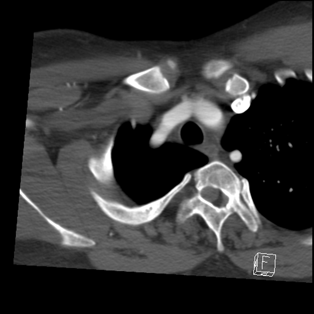
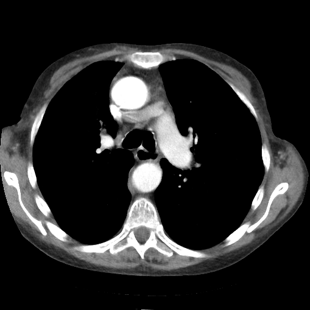
The retro-aortic left brachiocephalic vein is a rare vascular variant where the left brachiocephalic vein passes more inferiorly through the superior mediastinum, coursing inferior to the aortic arch and posterior to the ascending aorta to join the right brachiocepahilc vein forming the superior vena cava. The usual anatomy is for the vein to pass higher and more horizontally through the superior mediastinum, anterior to the branches of the aortic arch.
Associations
Retro-aortic left brachiocephalic vein has a high association with congenital cardiac disease, with the highest associations including:
References
- 1. Kulkarni S, Jain S, Kasar P, Garekar S, Joshi S. Retroaortic Left Innominate Vein - Incidence, Association with Congenital Heart Defects, Embryology, and Clinical Significance. Ann Pediatr Cardiol. 2008;1(2):139-41. doi:10.4103/0974-2069.43881 - Pubmed
- 2. Renan Uflacker. Atlas of Vascular Anatomy. (2007) ISBN: 9780781760812 - Google Books
- 3. Semionov A & Kosiuk J. Incidental Retroaortic Left Innominate Vein in Adult Patient. Radiol Case Rep. 2017;12(3):475-8. doi:10.1016/j.radcr.2017.05.005 - Pubmed
Incoming Links
Related articles: Anatomy: Thoracic
- thoracic skeleton[+][+]
- thoracic cage
- thoracic spine
- articulations
- muscles of the thorax[+][+]
- diaphragm
- intercostal space
- intercostal muscles
- variant anatomy
- spaces of the thorax[+][+]
- thoracic viscera[+][+]
- lower respiratory tract
-
heart
- cardiac chambers
- heart valves
- cardiac fibrous skeleton
- innervation of the heart
- development of the heart
- cardiac wall
-
pericardium
- epicardium
- epicardial fat pad
- pericardial space
- oblique pericardial sinus
- transverse pericardial sinus
-
pericardial recesses
- aortic recesses
- pulmonic recesses
- postcaval recess
- pulmonary venous recesses
- pericardial ligaments
- myocardium
- endocardium
-
pericardium
- esophagus
- thymus
- breast
- arterial supply of the thorax[+][+]
-
thoracic aorta (development)
-
ascending aorta
-
aortic root
- aortic annulus
-
coronary arteries
- coronary arterial dominance
- myocardial segments
-
left main coronary artery (LMCA)
- ramus intermedius artery (RI)
-
circumflex artery (LCx)
- obtuse marginal branches (OM1, OM2, etc))
- Kugel's artery
-
left anterior descending artery (LAD)
- diagonal branches (D1, D2, etc)
- septal perforators (S1, S2, etc)
-
right coronary artery (RCA)
- conus artery
- sinoatrial nodal artery
- acute marginal branches (AM1, AM2, etc)
- inferior interventricular artery (PDA)
- posterior left ventricular artery (PLV)
- congenital anomalies
- sinotubular junction
-
aortic root
- aortic arch
- aortic isthmus
- descending aorta
-
ascending aorta
- pulmonary trunk
-
thoracic aorta (development)
- venous drainage of the thorax
- superior vena cava (SVC)
- inferior vena cava (IVC)[+][+]
-
coronary veins[+][+]
-
cardiac veins which drain into the coronary sinus
- great cardiac vein
- middle cardiac vein
- small cardiac vein
- posterior vein of the left ventricle
- vein of Marshall (oblique vein of the left atrium)
- anterior cardiac veins
- venae cordis minimae (smallest cardiac veins or thebesian veins)
-
cardiac veins which drain into the coronary sinus
- pulmonary veins
- bronchial veins
- thoracoepigastric vein
- lymphatics of the thorax[+][+]
- innervation of the thorax[+][+]


 Unable to process the form. Check for errors and try again.