
Transversus thoracis muscle
Citation, DOI, disclosures and article data
At the time the article was created James Ling had no recorded disclosures.
View James Ling's current disclosuresAt the time the article was last revised Craig Hacking had no recorded disclosures.
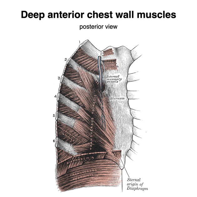
View Craig Hacking's current disclosuresThe transversus thoracis muscle is the innermost muscle of the anterior thoracic wall (deep to external intercostal and internal intercostal muscles).
Gross anatomy
The transversus thoracis is a thin band of muscle and tendon arising from the lower posterior surface of the sternum, posterior edge of the xiphisternum and the costal cartilage of the lowest 3-4 ribs at their sternal end. It diverges supero-laterally to attach by slips to the underside of the 2nd to 6th costal cartilages 1. The transversus thoracis muscle is morphologically identical to the transversus abdominis muscle 2.
Blood supply
Nerve supply
Action
The transversus thoracis muscle is an accessory muscle of respiration. It depresses the ribs which supports expiration.
References
- 1. Gray Henry, T. Pickering Pick and Robert Howden. Anatomy, descriptive and surgical. 39th ed. New York: Bounty Books, 1977
- 2. Sinnatamby, C. and Last, R. Last's anatomy. 12th ed. Edinburgh: Churchill Livingstone/Elsevier, 2011
- 3. West J. Respiratory physiology - the essentials. 9th ed. Philadelphia: Wolters Klower/Lippincott Williams and Wilkins, 2011
Incoming Links
Related articles: Anatomy: Thoracic
- thoracic skeleton[+][+]
- thoracic cage
- thoracic spine
- articulations
- muscles of the thorax
- diaphragm[+][+]
- intercostal space
-
intercostal muscles
- external intercostal muscle
- internal intercostal muscle
- innermost intercostal muscles
- subcostal muscles
- transversus thoracis muscle
- variant anatomy[+][+]
- spaces of the thorax[+][+]
- thoracic viscera[+][+]
- lower respiratory tract
-
heart
- cardiac chambers
- heart valves
- cardiac fibrous skeleton
- innervation of the heart
- development of the heart
- cardiac wall
-
pericardium
- epicardium
- epicardial fat pad
- pericardial space
- oblique pericardial sinus
- transverse pericardial sinus
-
pericardial recesses
- aortic recesses
- pulmonic recesses
- postcaval recess
- pulmonary venous recesses
- pericardial ligaments
- myocardium
- endocardium
-
pericardium
- esophagus
- thymus
- breast
- arterial supply of the thorax[+][+]
-
thoracic aorta (development)
-
ascending aorta
-
aortic root
- aortic annulus
-
coronary arteries
- coronary arterial dominance
- myocardial segments
-
left main coronary artery (LMCA)
- ramus intermedius artery (RI)
-
circumflex artery (LCx)
- obtuse marginal branches (OM1, OM2, etc))
- Kugel's artery
-
left anterior descending artery (LAD)
- diagonal branches (D1, D2, etc)
- septal perforators (S1, S2, etc)
-
right coronary artery (RCA)
- conus artery
- sinoatrial nodal artery
- acute marginal branches (AM1, AM2, etc)
- inferior interventricular artery (PDA)
- posterior left ventricular artery (PLV)
- congenital anomalies
- sinotubular junction
-
aortic root
- aortic arch
- aortic isthmus
- descending aorta
-
ascending aorta
- pulmonary trunk
-
thoracic aorta (development)
- venous drainage of the thorax[+][+]
- superior vena cava (SVC)
- inferior vena cava (IVC)
-
coronary veins
-
cardiac veins which drain into the coronary sinus
- great cardiac vein
- middle cardiac vein
- small cardiac vein
- posterior vein of the left ventricle
- vein of Marshall (oblique vein of the left atrium)
- anterior cardiac veins
- venae cordis minimae (smallest cardiac veins or thebesian veins)
-
cardiac veins which drain into the coronary sinus
- pulmonary veins
- bronchial veins
- thoracoepigastric vein
- lymphatics of the thorax[+][+]
- innervation of the thorax[+][+]


 Unable to process the form. Check for errors and try again.