
Thoracic aorta
Citation, DOI, disclosures and article data
At the time the article was created Jeremy Jones had no recorded disclosures.
View Jeremy Jones's current disclosuresAt the time the article was last revised Jeremy Jones had no financial relationships to ineligible companies to disclose.
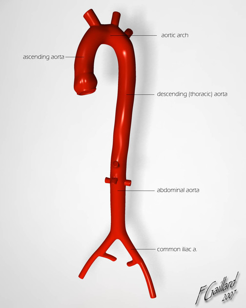
View Jeremy Jones's current disclosuresThe thoracic aorta is the most superior division of the aorta and is divided into three sections:
The thoracic aorta begins at the aortic valve, located obliquely just to the left of the midline at the level of the third intercostal space. It terminates as it exits the thorax to enter the abdomen through the median arcuate ligament between the diaphragmatic crurae anterior to the T12 vertebral body.
It supplies blood to the chest, upper limbs, head, and neck.
Typical measurements for adults (there can be considerable variation dependent on age):
aortic annulus: ~23 mm
aortic valve sinus / sinus of Valsalva: ~30 ± 5 mm
ascending aorta: 31 ± 4 mm
proximal to the brachiocephalic trunk: ~29 ± 4 mm
proximal transverse arch: ~28 ± 4 mm
distal transverse arch: ~26 ± 4 mm
aortic isthmus: ~25 ± 4 mm
at the diaphragm: ~24 ± 4 mm
Quiz questions
References
- 1. Last's Anatomy - 10th Edition - Chummy S Sinnatamby
- 2. Clemete's Anatomy - Regional Atlas of the Human Body - 3rd Edition
- 3. Gray's Anatomy 39th Edition, Elsevier
- 4. Keith L. Moore, Arthur F. Dalley, A. M. R. Agur. Clinically Oriented Anatomy. (2013) ISBN: 9781451119459 - Google Books
- 5. Freeman L, Young P, Foley T, Williamson E, Bruce C, Greason K. CT and MRI Assessment of the Aortic Root and Ascending Aorta. AJR Am J Roentgenol. 2013;200(6):W581-92. doi:10.2214/AJR.12.9531 - Pubmed
Incoming Links
- Thoracic aortic injury
- Bronchial artery
- Levator costarum muscle
- Ductus diverticulum
- Intercostal spaces
- Penetrating atherosclerotic ulcer
- Thoracic aortic aneurysm
- Superior phrenic arteries
- Windsock sign (aortic dissection)
- Aortic spindle
- Potts shunt
- Multisystemic smooth muscle dysfunction syndrome
- Esophagus
- Oesophageal bronchus
- Aorta
- Thorax
- Left ventricular outflow tract obstruction in echocardiography (differential)
- Interspinales muscles
- Coral reef aorta
- Left main bronchus
- Thoracic aortic ectasia with mural thrombus
- Massive aortic aneurysm, dissection and pericardial effusion
- Thoracic aorta - normal (transthoracic echocardiography)
- Thoracic aorta (illustration)
- Thoracic aortic injury (illustration)
- Extrapleural fat sign
- Aortic transection
- Right sided aortic arch with mirror image branching and absent coeliac trunk
- Thoracic aortic aneurysm
Related articles: Anatomy: Thoracic
- thoracic skeleton[+][+]
- thoracic cage
- thoracic spine
- articulations
- muscles of the thorax[+][+]
- diaphragm
- intercostal space
- intercostal muscles
- variant anatomy
- spaces of the thorax[+][+]
- thoracic viscera[+][+]
- lower respiratory tract
-
heart
- cardiac chambers
- heart valves
- cardiac fibrous skeleton
- innervation of the heart
- development of the heart
- cardiac wall
-
pericardium
- epicardium
- epicardial fat pad
- pericardial space
- oblique pericardial sinus
- transverse pericardial sinus
-
pericardial recesses
- aortic recesses
- pulmonic recesses
- postcaval recess
- pulmonary venous recesses
- pericardial ligaments
- myocardium
- endocardium
-
pericardium
- esophagus
- thymus
- breast
- arterial supply of the thorax
-
thoracic aorta (development)
-
ascending aorta
-
aortic root[+][+]
- aortic annulus
-
coronary arteries
- coronary arterial dominance
- myocardial segments
-
left main coronary artery (LMCA)
- ramus intermedius artery (RI)
-
circumflex artery (LCx)
- obtuse marginal branches (OM1, OM2, etc))
- Kugel's artery
-
left anterior descending artery (LAD)
- diagonal branches (D1, D2, etc)
- septal perforators (S1, S2, etc)
-
right coronary artery (RCA)
- conus artery
- sinoatrial nodal artery
- acute marginal branches (AM1, AM2, etc)
- inferior interventricular artery (PDA)
- posterior left ventricular artery (PLV)
- congenital anomalies
- sinotubular junction
-
aortic root[+][+]
- aortic arch
- aortic isthmus
- descending aorta
-
ascending aorta
- pulmonary trunk[+][+]
-
thoracic aorta (development)
- venous drainage of the thorax[+][+]
- superior vena cava (SVC)
- inferior vena cava (IVC)
-
coronary veins
-
cardiac veins which drain into the coronary sinus
- great cardiac vein
- middle cardiac vein
- small cardiac vein
- posterior vein of the left ventricle
- vein of Marshall (oblique vein of the left atrium)
- anterior cardiac veins
- venae cordis minimae (smallest cardiac veins or thebesian veins)
-
cardiac veins which drain into the coronary sinus
- pulmonary veins
- bronchial veins
- thoracoepigastric vein
- lymphatics of the thorax[+][+]
- innervation of the thorax[+][+]


 Unable to process the form. Check for errors and try again.