
Esophageal bronchus
Citation, DOI, disclosures and article data
Citation:
Knipe H, Saber M, Debowski M, et al. Esophageal bronchus. Reference article, Radiopaedia.org (Accessed on 26 Mar 2025) https://doi.org/10.53347/rID-29810
rID:
29810
Article created:
Disclosures:
At the time the article was created Henry Knipe had no recorded disclosures.
View Henry Knipe's current disclosures
Last revised:
Disclosures:
At the time the article was last revised Mohamed Saber had no recorded disclosures.
View Mohamed Saber's current disclosures
Revisions:
5 times, by
4 contributors -
see full revision history and disclosures
Systems:
Sections:
Tags:
Synonyms:
- Esophageal bronchus
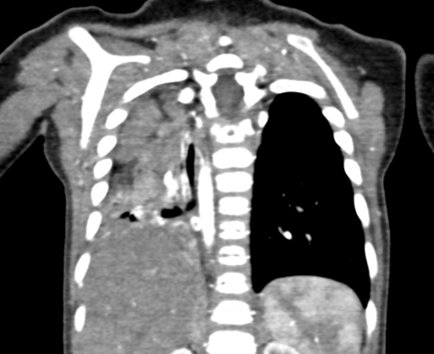
Esophageal bronchus, a.k.a. communicating bronchopulmonary foregut malformation, refers to the rare occurrence where a bronchus arises directly from the esophagus.
On this page:
Epidemiology
It is more common in females with a M:F of 1:2 2.
Gross anatomy
Esophageal bronchi may be the main bronchus, which gives rise to esophageal lung, or may be a lobar bronchus, most commonly a lower lobe bronchus.
Associations
Blood supply
Arterial supply and venous drainage are variable:
- arterial supply: may arise directly from the thoracic aorta
- venous drainage: may be to systemic or pulmonary circulations
Radiographic appearance
Plain radiograph
- unilateral alveolar opacity and air bronchogram as a result of aspiration 4
- mediastinal shift to the affected side 2
Fluoroscopy
- barium fills the bronchus and first-order branches 2
References
- 1. Stringer DA, Babyn PS. Pediatric Gastrointestinal Imaging and Intervention. B. C. Decker. ISBN:1550090798. Read it at Google Books - Find it at Amazon
- 2. Lallemand D, Quignodon J, Courtel J. Pediatric Radiology. 1996;26 (3): . doi:10.1007/BF01405293
- 3. Coley BD. Caffey's Pediatric Diagnostic Imaging (Caffeys Pediatric Diagnostic Imaging). Saunders. ISBN:B00D5T5MMY. Read it at Google Books - Find it at Amazon
Incoming Links
Related articles: Anatomy: Thoracic
- thoracic skeleton[+][+]
- thoracic cage
- thoracic spine
- articulations
- muscles of the thorax[+][+]
- diaphragm
- intercostal space
- intercostal muscles
- variant anatomy
- spaces of the thorax[+][+]
- thoracic viscera
-
lower respiratory tract
- tracheobronchial tree
-
lungs[+][+]
-
bronchopulmonary segmental anatomy (Boyden Classification) (mnemonic)
- left lung
- right lung
- variant anatomy
- lung parenchyma
- hilum
- pleura
-
bronchopulmonary segmental anatomy (Boyden Classification) (mnemonic)
-
heart[+][+]
- cardiac chambers
- heart valves
- cardiac fibrous skeleton
- innervation of the heart
- development of the heart
- cardiac wall
-
pericardium
- epicardium
- epicardial fat pad
- pericardial space
- oblique pericardial sinus
- transverse pericardial sinus
-
pericardial recesses
- aortic recesses
- pulmonic recesses
- postcaval recess
- pulmonary venous recesses
- pericardial ligaments
- myocardium
- endocardium
-
pericardium
- esophagus[+][+]
- thymus[+][+]
- breast[+][+]
-
lower respiratory tract
- arterial supply of the thorax[+][+]
-
thoracic aorta (development)
-
ascending aorta
-
aortic root
- aortic annulus
-
coronary arteries
- coronary arterial dominance
- myocardial segments
-
left main coronary artery (LMCA)
- ramus intermedius artery (RI)
-
circumflex artery (LCx)
- obtuse marginal branches (OM1, OM2, etc))
- Kugel's artery
-
left anterior descending artery (LAD)
- diagonal branches (D1, D2, etc)
- septal perforators (S1, S2, etc)
-
right coronary artery (RCA)
- conus artery
- sinoatrial nodal artery
- acute marginal branches (AM1, AM2, etc)
- inferior interventricular artery (PDA)
- posterior left ventricular artery (PLV)
- congenital anomalies
- sinotubular junction
-
aortic root
- aortic arch
- aortic isthmus
- descending aorta
-
ascending aorta
- pulmonary trunk
-
thoracic aorta (development)
- venous drainage of the thorax[+][+]
- superior vena cava (SVC)
- inferior vena cava (IVC)
-
coronary veins
-
cardiac veins which drain into the coronary sinus
- great cardiac vein
- middle cardiac vein
- small cardiac vein
- posterior vein of the left ventricle
- vein of Marshall (oblique vein of the left atrium)
- anterior cardiac veins
- venae cordis minimae (smallest cardiac veins or thebesian veins)
-
cardiac veins which drain into the coronary sinus
- pulmonary veins
- bronchial veins
- thoracoepigastric vein
- lymphatics of the thorax[+][+]
- innervation of the thorax[+][+]


 Unable to process the form. Check for errors and try again.