The tracheobronchial tree is the branching tree of airways beginning at the larynx and extending inferiorly and peripherally into the lungs as bronchioles. The luminal diameter decreases as the branching increases more peripherally into the lungs. The airway walls down to the level of the bronchioles contain C-shaped rings composed of hyaline cartilage to maintain luminal patency.
On this page:
Terminology
large airways: trachea and main, lobar, and segmental bronchi 4
small airways: subsegmental bronchi to bronchioles 4
Gross anatomy
When described as an inverted tree, the tracheobronchial tree constitutes:
the trunk: the trachea
two major branches: the left and right main (primary) bronchi, which bifurcate at the carina located in the transthoracic plane of Ludwig
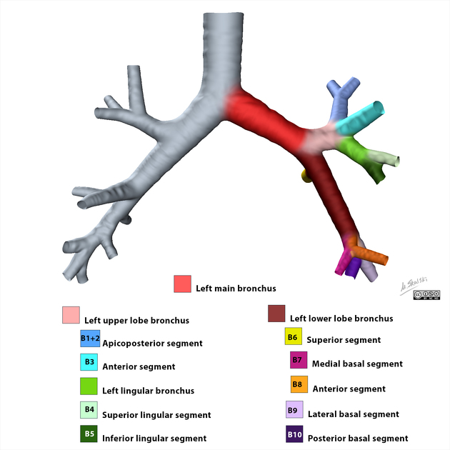
several intermediate branches: the lobar (secondary) and segmental (tertiary) bronchi within the lungs
many further consistent smaller and smaller subdivisions, terminating in the alveoli in the peripheral of the lungs
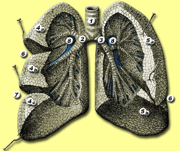
At the level of the tertiary segmental bronchi, sections of the lung called bronchopulmonary segments are defined as representing the largest subdivisions of a lobe that can be surgically resectable, analogous to the segments of the liver. Each bronchopulmonary segment is separated from adjacent segments by thin fibrous septa (pulmonary interstitium) and is pyramidal-shaped with the apex of the pyramid facing the lung root and the base of the pyramid facing the pleural surface.
Bronchopulmonary segments are independently supplied by a single segmental bronchus and a tertiary branch of the pulmonary artery, which both enter the segment at its apex. Venous drainage occurs through intersegmental tributaries of the pulmonary veins, located in the interstitium between segments.
Peripheral to the segmental bronchi there are 20-25 further branchings into conducting bronchioles, and finally terminal bronchioles. These structures lack cartilage and do not participate in gas exchange. Their function is to transport air to the most distal parts of the lungs where respiratory bronchioles are located.
Respiratory bronchioles contain numerous small thin-walled alveoli that bulge outwards from their lumens. As they contain alveoli, respiratory bronchioles are thus involved in both gas exchange and air transportation.
Further subdivision occurs beyond the respiratory bronchioles, to alveolar ducts and alveolar sacs. Lastly, alveoli are the single basic structural unit responsible for gas exchange in the lung. There are approximately 300 million alveoli in the lungs of normal young adults.
Arterial supply
The trachea is supplied by tracheal arteries, branches of the inferior thyroid artery and bronchial arteries. The tracheobronchial tree from the carina to the respiratory bronchioles is supplied by bronchial arteries, branches of the descending aorta. There are usually two bronchial arteries on the left and a single bronchial artery on the right.
Venous drainage
Venous drainage occurs via the bronchial veins which accompany the bronchi and drain into the azygos system on the right and the accessory hemiazygos vein on the left. Deeper within the lung parenchymal, venous blood will drain into pulmonary veins thus mixing with oxygenated blood in the left atrium.
Lymphatic drainage
The tracheobronchial tree is drained by lymphatic channels which course along the bronchi and pulmonary arteries towards the hilum. Ultimately these drain to hilar lymph nodes and then ascend to the mediastinal lymph nodes.


 Unable to process the form. Check for errors and try again.