
The aortic valve (AV) is one of the four cardiac valves and one of two semilunar valves (along with the pulmonary valve). It allows blood to exit the left ventricle (LV) during systole by opening, and during diastole it stops blood exiting by closing.
On this page:
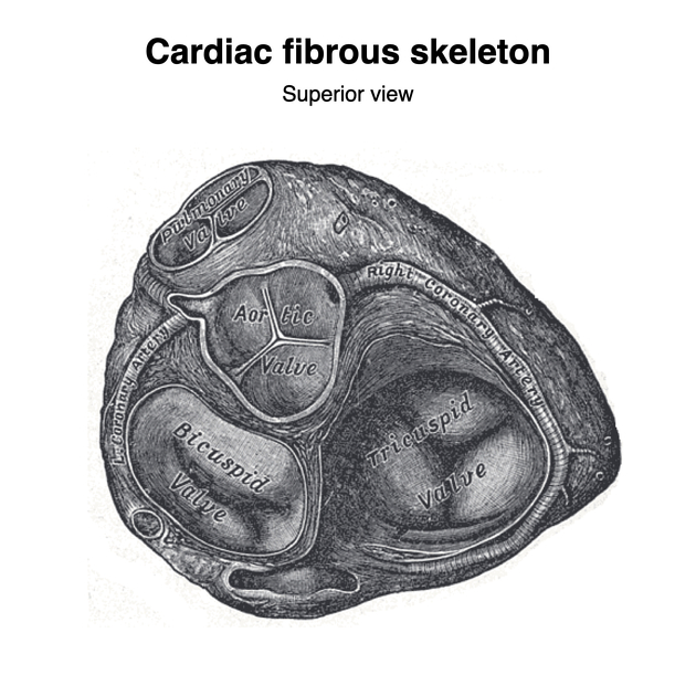
Gross anatomy
The valve has left, right, and posterior cusps, the bases of which attach around the valve orifice to a fibrous ring or annulus, forming part of the fibrous skeleton of the heart. The cusps attach to each other and to the annulus at the commissures.
The free edge of each cusp (the lunule) is thickened where it contacts the free edges of adjacent cusps and at the angulated apex of each free edge, there is further nodular thickening, known as the nodule (of Arantius). The nodules should touch each other during diastole with the valve fully closed. The cusps bulge inferiorly into the outflow tract of the left ventricle.
Immediately superior to the cusps, the ascending aorta is mildly dilated, forming the aortic sinuses, the spaces between the dilated wall of the aorta and the cusps of the semilunar valve. During systole, these sinuses prevent the cusps from flattening against the walls of the sinuses, which may restrict valve closure during diastole.
The left coronary artery arises from the left aortic sinus and the right coronary artery arises from the right aortic sinus. The posterior sinus does not have a vessel arising from it and is, therefore, referred to as the non-coronary sinus. The aortic valve lies obliquely and is inferior and to the right of the pulmonary valve (PV).
Variant anatomy
unicuspid aortic valve
Related pathology


 Unable to process the form. Check for errors and try again.