
The myocardium is the middle layer of the cardiac wall between the endocardium and the pericardium and forms the muscular part of the heart.
On this page:
Gross anatomy
The myocardium represents the middle layer of the cardiac wall. It is located between the endocardium and the epicardial layer of the pericardium within the walls of the cardiac chambers arranged in different sheets wrapped around in different orientations.
The left ventricular myocardium can be subdivided into the following layers or zones from inside to outside:
subendocardial
midmyocardial
subepicardial
The exact myocardial architecture is somewhat controversial. It has been compared to a contractile complex three-dimensional mesh, made up of myocytes merging with their neighbouring cells in a nonuniform anisotropic manner supported by an extracellular collagenous matrix-forming bundle of myofibres, which are configured in a helical or spiral pattern within the two ventricles 1-4.
The ventricular myocardium is thicker than the atrial myocardium, in particular, the myocardium of the left ventricle.
Boundaries
The inner border merges with the subendocardial tissue layer, which contains collagen, elastic fibres small blood vessels and nerves as well as the Purkinje fibres and connects the extracellular matrix of the myocardium to the endocardium 4.
The outer border is formed by the subepicardial layer, which is interconnected to the epicardium, the latter consisting of a lining of mesothelial cells, a subserosal layer of connective tissue and a variable amount of epicardial fatty tissue.
Arterial supply
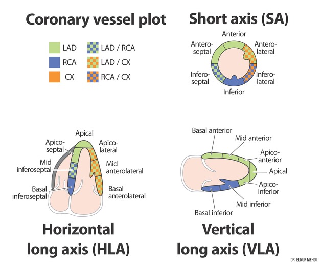
The arterial supply of the myocardium is provided by the coronary arteries.
The vascular territories of the left ventricular myocardium are divided into 17 segments and illustrated by the cardiac segmentation model of the American Heart Association (AHA) 6,7.
Venous drainage
Venous drainage of the myocardium is provided via the cardiac veins and the coronary sinus as well as the Thebesian veins 8.
Lymphatic drainage
Cardiac lymphatic flow passes from the endocardium through the myocardium to the epicardium where small lymphatic vessels drain into larger collecting vessels. In this process, myocardial contractions help to advance lymph flow. The lymphatic function is critical to maintaining the myocardial interstitial fluid equilibrium and cardiac function and it has been advocated that it aids tissue repair and prevents adverse remodelling in case of myocardial injury 9.
Innervation
The myocardium is innervated by the cardiac conduction system. Myocardial conduction happens from cardiomyocyte to cardiomyocyte via the intercalated disks, which form the mechanical and electrical contacts between the myocardial cells 10.
Histology
The myocardium consists of cardiomyocytes grouped in strands also known as myofibres and the surrounding extracellular matrix with endomysial and perimysial components. The cardiomyocytes are linked with each other through distinctive junctional compounds, the intercalated discs, which facilitate intercellular electrical impulse conduction. The myocyte strands diverge in different angles from the lining of the epicardial surface 1-5.
Radiographic features
The myocardium can be depicted and evaluated with ultrasound i.e. echocardiography, cardiac CT and cardiac MRI.
In most imaging techniques the myocardium displays a muscle-like appearance.
Ultrasound
Echocardiography
Echocardiography has been traditionally used as a first-line imaging technique in the evaluation of cardiac morphology and function as well as in the assessment of myocardial contractility and the contractile reserve 11.
It has been also used for the evaluation of myocardial strain or simplified for the assessment of regional deformations of the myocardium such as thickening, shortening or lengthening. Echocardiographic strain imaging techniques include tissue Doppler imaging and speckle tracking 12.
CT
Cardiac CT can be used for the assessment of myocardial morphology and myocardial perfusion. In addition, research has been conducted concerning the use as an alternative for myocardial extracellular volume quantification 13,14.
MRI
The myocardium can be visualised and is routinely evaluated with a whole series of sequences and imaging techniques assessing its function and inherent tissue properties, such as 15-18:
cine imaging: wall morphology, contractile function, myocardial strain (myocardial feature tracking)
black blood imaging: wall morphology and tissue properties such as myocardial oedema
-
myocardial mapping: inherent T1, T2 and T2* values as well as extracellular volume assessment
T2 mapping: myocardial oedema
T2* mapping: iron deposition, myocardial haemorrhage
ECV: myocardial oedema/myocardial fibrosis
myocardial perfusion imaging: myocardial perfusion
late gadolinium enhancement: changes in distribution patterns of gadolinium-based contrast agents within the myocardium
Specific less often used cardiac imaging techniques for the evaluation of the myocardium include:
myocardial tagging: contractile function, myocardial strain
arterial spin labelling (ASL): myocardial perfusion
In addition, the myocardial architecture and myofibril ultrastructure has been investigated with diffusion tensor imaging. Due to long acquisition times and this imaging technique has been mainly used in research 3.
Nuclear medicine
SPECT/PET
SPECT and PET imaging are used in myocardial perfusion imaging and the assessment of myocardial viability. PET permits the measurement of absolute myocardial blood flow. Furthermore, it can be used in the evaluation of myocardial inflammation 19.
History and etymology
The word myocardium is derived from the Greek words 'myo-' muscle and 'kardia' heart.
Related pathology
The following pathologies and diseases are related to the myocardium:
-
myocardial inflammation/myocarditis
-
cardiomyopathies
and many more...


 Unable to process the form. Check for errors and try again.