
Aortic root
Citation, DOI, disclosures and article data
At the time the article was created Laura Smith had no recorded disclosures.
View Laura Smith's current disclosuresAt the time the article was last revised Jeremy Jones had no financial relationships to ineligible companies to disclose.
View Jeremy Jones's current disclosures- Aortic root anatomy
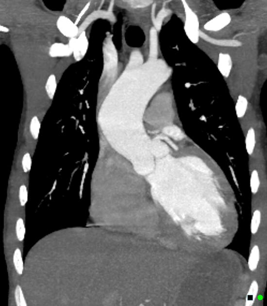
The aortic root is the first part of the aorta containing parts of the aortic valve and connects the heart to systemic circulation.
Gross anatomy
The aortic root is located between the aortic annulus (the junction of the outflow tract of the left ventricle and the aortic valve) and the sinotubular junction (where the ascending aorta originates).
The average aortic diameter at the sinuses of Valsalva is often taken as 3.0 cm ± 0.5 4.
It has several subparts 1:
three aortic valve leaflets and leaflet attachments
-
three aortic sinuses of Valsalva: intraluminal space formed by the cusps
right and left contain coronary aorta ostia, with a third non-coronary aortic sinus located posteriorly
three interleaflet trigones: space between the cusps and the annulus
sinotubular junction, which divides the aortic root from the ascending aorta
Relations
anterior: ascending aorta
posterior: left ventricle
medial: right coronary artery
lateral: left coronary artery
Related pathology
References
- 1. Charitos E & Sievers H. Anatomy of the Aortic Root: Implications for Valve-Sparing Surgery. Ann Cardiothorac Surg. 2013;2(1):53-6. doi:10.3978/j.issn.2225-319X.2012.11.18 - Pubmed
- 2. Goldstein S, Evangelista A, Abbara S et al. Multimodality Imaging of Diseases of the Thoracic Aorta in Adults: From the American Society of Echocardiography and the European Association of Cardiovascular Imaging: Endorsed by the Society of Cardiovascular Computed Tomography and Society for Cardiovascular Magnetic Resonance. J Am Soc Echocardiogr. 2015;28(2):119-82. doi:10.1016/j.echo.2014.11.015 - Pubmed
- 3. Freeman L, Young P, Foley T, Williamson E, Bruce C, Greason K. CT and MRI Assessment of the Aortic Root and Ascending Aorta. AJR Am J Roentgenol. 2013;200(6):W581-92. doi:10.2214/AJR.12.9531 - Pubmed
- 4. Hanneman K, Chan F, Mitchell R, Miller D, Fleischmann D. Pre- and Postoperative Imaging of the Aortic Root. Radiographics. 2016;36(1):19-37. doi:10.1148/rg.2016150053 - Pubmed
- 5. Underwood M, El Khoury G, Deronck D, Glineur D, Dion R. The Aortic Root: Structure, Function, and Surgical Reconstruction. Heart. 2000;83(4):376-80. doi:10.1136/heart.83.4.376 - Pubmed
- 6. Ko J, Goldstein J, Latson L et al. Chest CT Angiography for Acute Aortic Pathologic Conditions: Pearls and Pitfalls. Radiographics. 2021;41(2):399-424. doi:10.1148/rg.2021200055 - Pubmed
- 7. Charitos E & Sievers H. Anatomy of the Aortic Root: Implications for Valve-Sparing Surgery. Ann Cardiothorac Surg. 2013;2(1):53-6. doi:10.3978/j.issn.2225-319X.2012.11.18 - Pubmed
- 8. Tretter J, Izawa Y, Spicer D et al. Understanding the Aortic Root Using Computed Tomographic Assessment: A Potential Pathway to Improved Customized Surgical Repair. Circ Cardiovasc Imaging. 2021;14(11):e013134. doi:10.1161/CIRCIMAGING.121.013134 - Pubmed
Incoming Links
- Ascending aorta
- Hypertrophic cardiomyopathy protocol (MRI)
- Sinotubular junction
- Pulmonary artery intramural haematoma
- Hypoplastic left heart syndrome
- Congenital heart disease in echocardiography (an approach)
- Cardiac CT (an approach)
- Retroaortic coronary course
- Cardiac calcification
- Right atrium
- Sinus of Valsalva
- Aortic annulus
- Ross procedure
- Aortic root to right ventricle fistula
- Kugel artery
- Mitral annulus
- Acute aortic syndrome
- Aortic root dilatation
- Atrial septum
- Annuloaortic ectasia
Related articles: Anatomy: Thoracic
- thoracic skeleton[+][+]
- thoracic cage
- thoracic spine
- articulations
- muscles of the thorax[+][+]
- diaphragm
- intercostal space
- intercostal muscles
- variant anatomy
- spaces of the thorax[+][+]
- thoracic viscera[+][+]
- lower respiratory tract
-
heart
- cardiac chambers
- heart valves
- cardiac fibrous skeleton
- innervation of the heart
- development of the heart
- cardiac wall
-
pericardium
- epicardium
- epicardial fat pad
- pericardial space
- oblique pericardial sinus
- transverse pericardial sinus
-
pericardial recesses
- aortic recesses
- pulmonic recesses
- postcaval recess
- pulmonary venous recesses
- pericardial ligaments
- myocardium
- endocardium
-
pericardium
- esophagus
- thymus
- breast
- arterial supply of the thorax
-
thoracic aorta (development)
-
ascending aorta
-
aortic root
- aortic annulus
-
coronary arteries
- coronary arterial dominance
- myocardial segments
-
left main coronary artery (LMCA)[+][+]
- ramus intermedius artery (RI)
-
circumflex artery (LCx)
- obtuse marginal branches (OM1, OM2, etc))
- Kugel's artery
-
left anterior descending artery (LAD)
- diagonal branches (D1, D2, etc)
- septal perforators (S1, S2, etc)
-
right coronary artery (RCA)[+][+]
- conus artery
- sinoatrial nodal artery
- acute marginal branches (AM1, AM2, etc)
- inferior interventricular artery (PDA)
- posterior left ventricular artery (PLV)
- congenital anomalies
- sinotubular junction
-
aortic root
- aortic arch[+][+]
- aortic isthmus[+][+]
- descending aorta[+][+]
-
ascending aorta
- pulmonary trunk[+][+]
-
thoracic aorta (development)
- venous drainage of the thorax[+][+]
- superior vena cava (SVC)
- inferior vena cava (IVC)
-
coronary veins
-
cardiac veins which drain into the coronary sinus
- great cardiac vein
- middle cardiac vein
- small cardiac vein
- posterior vein of the left ventricle
- vein of Marshall (oblique vein of the left atrium)
- anterior cardiac veins
- venae cordis minimae (smallest cardiac veins or thebesian veins)
-
cardiac veins which drain into the coronary sinus
- pulmonary veins
- bronchial veins
- thoracoepigastric vein
- lymphatics of the thorax[+][+]
- innervation of the thorax[+][+]


 Unable to process the form. Check for errors and try again.