
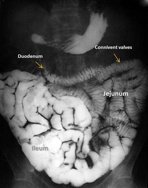
The duodenum (plural: duodena or duodenums) is the first part of the small intestine and is the continuation of the stomach.
On this page:
Gross anatomy
The duodenum is a 20-30 cm C-shaped hollow viscus predominantly on the right side of the vertebral column. It lies at the level of L1-3 and the convexity of the duodenum (called the duodenal sweep by radiologists) usually encompasses the head of the pancreas.
The duodenum begins at the duodenal bulb and ends at the ligament of Treitz, where it continues as the jejunum (this is often called the duodenojejunal (DJ) flexure). It is composed of four distinct parts. The first 2.5 cm of the first part of the duodenum is intraperitoneal. Meanwhile, the remaining parts of the duodenum are retroperitoneal. The anterior surface of duodenum is mostly covered by the peritoneum except for second part of the duodenum which is crossed by transverse mesocolon and the third part which is crossed by superior mesenteric vessels 5.
Segments
First part (D1)
The first (superior) part of the duodenum begins as a continuation of the duodenal end of the pylorus. It passes laterally to the right, superiorly and posteriorly, for approximately 5 cm, before making a sharp curve inferiorly into the superior duodenal flexure. It is intraperitoneal for the first 2-3 cm only. Relations 3:
anteriorly: gallbladder, liver
posteriorly: common bile duct, portal vein, gastroduodenal artery
superiorly: epiploic foramen
inferiorly: pancreatic head
Second part (D2)
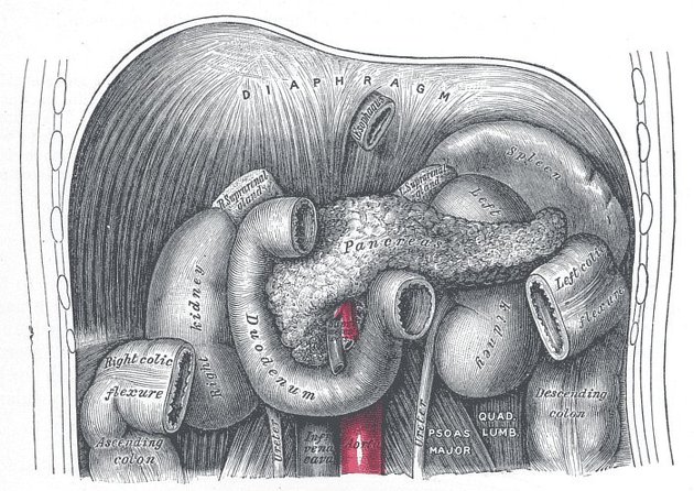
The second (descending) part of the duodenum begins at the superior duodenal flexure. It passes inferiorly to the lower border of vertebral body L3, before making a sharp turn medially into the inferior duodenal flexure, the end of the descending part. Relations 3:
anteriorly: transverse mesocolon
posteriorly: right kidney, right ureter, right adrenal gland
superiorly: liver, gallbladder (variable)
inferiorly: loops of jejunum
laterally: ascending colon, hepatic flexure, right kidney
medially: pancreatic head
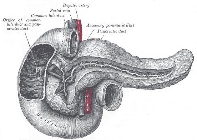
The pancreatic duct and common bile duct enter the descending duodenum through the major duodenal papilla (ampulla of Vater). This part of the duodenum also contains the minor duodenal papilla, the entrance for the accessory pancreatic duct. The junction between the embryological foregut and midgut lies just below the major duodenal papilla.
Third part (D3)
The third (inferior/horizontal) part of the duodenum begins at the inferior duodenal flexure and passes transversely to the left, crossing the vertebral column. Relations 3:
anteriorly: small bowel mesentery root
posteriorly: right psoas muscle, right ureter, gonadal vessels, aorta and IVC
superiorly: pancreatic head
inferiorly: loops of jejunum
Fourth part (D4)
The fourth (ascending) part passes superiorly, either anterior to, or to the left of, the aorta, until it reaches the inferior border of the body of the pancreas. Then, it curves anteriorly and terminates at the duodenojejunal flexure where it joins the jejunum. The duodenojejunal flexure is surrounded by a peritoneal fold containing muscle fibres: the ligament of Treitz. Relations 3:
superiorly: stomach
inferiorly: loops of jejunum
posteriorly: left psoas muscle, aorta
Arterial supply
duodenal cap (first 2.5 cm): right gastric artery, right gastroepiploic artery
remaining D1 to mid-D2: superior pancreaticoduodenal artery (branch of gastroduodenal artery)
mid-D2 to ligament of Treitz: inferior pancreaticoduodenal arteries (branch of SMA) 3
Venous drainage
duodenal cap (first 2.5 cm): prepyloric vein (drains to portal vein)
remaining duodenum: superior pancreaticoduodenal vein (drains to portal vein) and inferior pancreaticoduodenal vein (drains to superior mesenteric vein) 3
Lymphatic drainage
-
pancreaticoduodenal nodes that drain:
distally to superior mesenteric nodes
proximally to coeliac nodes 3
Innervation
sympathetic: via the coeliac plexus and superior mesenteric plexus
parasympathetic: via anterior and posterior vagal trunks 2
Variant anatomy
duodenal diverticulum: most commonly occurs in D2 or D3 4
duodenal duplication: most commonly occurs at the medial wall of D2 or D3; appears as a cystic structure that does not communicate with the lumen 4
History and etymology
Duodenum means "twelve" in Latin and the name derives from the belief that it measured twelve finger widths in length (duodenum digitorum) 4.


 Unable to process the form. Check for errors and try again.