
Manubriosternal joint
Citation, DOI, disclosures and article data
At the time the article was created James Ling had no recorded disclosures.
View James Ling's current disclosuresAt the time the article was last revised Henry Knipe had the following disclosures:
- Integral Diagnostics, Shareholder (ongoing)
- Micro-X Ltd, Shareholder (ongoing)
These were assessed during peer review and were determined to not be relevant to the changes that were made.
View Henry Knipe's current disclosures- Sternomanubrial joint
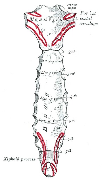
The manubriosternal joint, sometimes referred to as the sternomanubrial joint, is the articulation between the upper two parts of the sternum, the manubrium and sternal body. It is at the level of the sternal angle or angle of Louis, which is at the 2nd costal cartilage and the intervertebral disc of T4 and T5 1. This forms an important palpable landmark for clinical examination
Gross anatomy
The manubriosternal joint is a type of secondary cartilaginous joint or symphysis, formed by the inferior border of the manubrium and the superior border of the sternal body. Both sides of the joint are irregular and undulating and covered with hyaline cartilage 2. However, it is not a typical secondary cartilaginous joint as the bones may ossify later in adult life 3. The joint has an anterior and posterior ligament 4.
Function
There is very little movement of the manubriosternal joint but there may be a small amount of angular movement during respiration 5.
Related pathology
References
- 1. Keith L. Moore, Arthur F. Dalley, A. M. R. Agur. Clinically Oriented Anatomy. (2013) ISBN: 9781451119459 - Google Books
- 2. Chummy S. Sinnatamby. Last's Anatomy. (2011) ISBN: 9780702033957 - Google Books
- 3. VISHRAM. SINGH. Textbook of Anatomy (Regional and Clinical) Upper Limb and Thorax:. (2014) ISBN: 9788131237298 - Google Books
- 4. Cheney N, Taylor B, French B, Esterline W. Traumatic Sternomanubrial Instability and Arthrosis: A Case Report and Review. JBJS Case Connect. 2012;2(4):e67. doi:10.2106/JBJS.CC.L.00011 - Pubmed
- 5. Henry Gray, Patricia Collins. Gray's Anatomy. (2005) ISBN: 9780443071683 - Google Books
Incoming Links
Related articles: Anatomy: Thoracic
- thoracic skeleton
- thoracic cage[+][+]
- thoracic spine
- articulations
- sternoclavicular joint
- manubriosternal joint
- costochondral joint
- interchondral joint
- costovertebral joint
- xiphisternal joint
- muscles of the thorax[+][+]
- diaphragm
- intercostal space
- intercostal muscles
- variant anatomy
- spaces of the thorax[+][+]
- thoracic viscera[+][+]
- lower respiratory tract
-
heart
- cardiac chambers
- heart valves
- cardiac fibrous skeleton
- innervation of the heart
- development of the heart
- cardiac wall
-
pericardium
- epicardium
- epicardial fat pad
- pericardial space
- oblique pericardial sinus
- transverse pericardial sinus
-
pericardial recesses
- aortic recesses
- pulmonic recesses
- postcaval recess
- pulmonary venous recesses
- pericardial ligaments
- myocardium
- endocardium
-
pericardium
- esophagus
- thymus
- breast
- arterial supply of the thorax[+][+]
-
thoracic aorta (development)
-
ascending aorta
-
aortic root
- aortic annulus
-
coronary arteries
- coronary arterial dominance
- myocardial segments
-
left main coronary artery (LMCA)
- ramus intermedius artery (RI)
-
circumflex artery (LCx)
- obtuse marginal branches (OM1, OM2, etc))
- Kugel's artery
-
left anterior descending artery (LAD)
- diagonal branches (D1, D2, etc)
- septal perforators (S1, S2, etc)
-
right coronary artery (RCA)
- conus artery
- sinoatrial nodal artery
- acute marginal branches (AM1, AM2, etc)
- inferior interventricular artery (PDA)
- posterior left ventricular artery (PLV)
- congenital anomalies
- sinotubular junction
-
aortic root
- aortic arch
- aortic isthmus
- descending aorta
-
ascending aorta
- pulmonary trunk
-
thoracic aorta (development)
- venous drainage of the thorax[+][+]
- superior vena cava (SVC)
- inferior vena cava (IVC)
-
coronary veins
-
cardiac veins which drain into the coronary sinus
- great cardiac vein
- middle cardiac vein
- small cardiac vein
- posterior vein of the left ventricle
- vein of Marshall (oblique vein of the left atrium)
- anterior cardiac veins
- venae cordis minimae (smallest cardiac veins or thebesian veins)
-
cardiac veins which drain into the coronary sinus
- pulmonary veins
- bronchial veins
- thoracoepigastric vein
- lymphatics of the thorax[+][+]
- innervation of the thorax[+][+]


 Unable to process the form. Check for errors and try again.