
The breast is an apocrine gland found in both males and females. However, in females, it has a specific function - the production of milk for neonatal nutrition and immune function.
On this page:
Gross anatomy
The breast has an inhomogeneous structure that is predominantly composed of adipose tissue and glandular tissue. In addition, there are also suspensory Cooper's ligaments and connective tissue such as collagen and elastin.
The adult breast has nearly 14-18 lactiferous lobes. Each lobe is made up of several lobules, and each lobule is made up of several acini. Each acinus drains into a branching duct that converges into a single lactiferous duct in each lobe, that subsequently drain at the nipple-areola complex. The breast parenchyma comprised of glandular structures namely acini and ductal tissue. Meanwhile, the breast stroma comprised of fat and fibrous tissue 4.
The glandular parenchyma is estrogen dependent. During adolescence, the growing breast becomes increasingly glandular. During pregnancy and lactation, the number of acini increases. When lactation stops, the becomes even less glandular when compared to before pregnancy. Thus, the breast of a parous woman is less glandular when compared to a nulliparous woman of the same age 4.
Breast parenchyma starts to atrophy in adulthood and accelerated in menopause with increasing amount of fat 4.
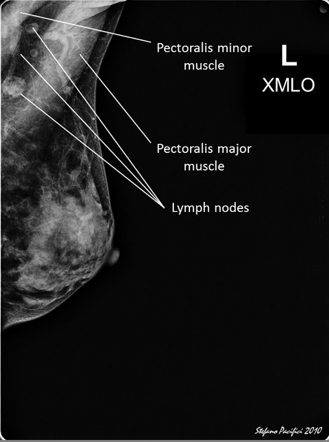
Relations
The breasts overlie the pectoralis major muscles and extend from the level of the 2nd to 6th ribs:
superior: clavicle
inferior: middle of the sternum
lateral: midaxillary line
medial: sternum
There is often an extension of breast tissue into the axilla called the axillary tail.
Arterial supply
internal thoracic artery perforators (2nd to 5th)
vessels to serratus anterior
terminal branches of 3rd to 8th intercostal perforators
Venous drainage
Lymphatic drainage
The drainage of lymph from the breast has a significant impact on the spread of malignancy and as such, has a separate article: lymphatic drainage of the breast.
Innervation
thoracic intercostal nerve T3-T5
supraclavicular nerve from the cervical plexus
Variant anatomy
polythelia (supernumerary nipple)
polymastia (accessory breast tissue)
Radiographic features
Mammography
shows a wide variation of homogeneously dense, milky structures (representing glandular tissue) interrupted by areas of curved or round radiolucent fat
Cooper's ligaments appear as curved, linear radiopacities
the duct system is not normally visualized except near the nipple 2
See articles: mammography views; breast density.
Ultrasound
glandular tissue appears variably hyperechoic and fat appears hypoechoic 4
lactiferous ducts appear as regular interspersed tubular, anechoic structures 4
Cooper's ligaments appear as hyperechoic, linear structures and may cause acoustic shadowing 2,3
the nipple can also cause acoustic shadowing, sometimes creating a pseudomass 3
retromammary fat is anechoic 4
See article: breast ultrasound.
MRI
T1: fat has moderate-high signal; glandular, ductal and connective tissue has low signal. Normal ducts are usually not visible on MRI unless they are dilated. Accumulation of hemorrhagic or proteinaceous material within obstructed duct will produce high T1-weighted signal 6.
T2: fat has high signal while fibroglandular tissue has low signal 4
-
T1 C+ (Gd): normal breast tissue is mildy enhancing with progressive, low enhancement overtime. During the first and the fourth week of menstrual cycle, enhancement maybe more rapid, producing a diffuse or focal pattern. Nipple and areolar complex may be intensely enhanced 5.
See article: breast MRI.
Development
During embryological development, breast tissue first appears as ectoderm ridges during the 6th week of gestation. This ridge grows thicker and leads to mesodermal compression. With further proliferation of the ectodermal cells, there is a growth of the same into the mesodermal layer leading to a formation of clusters that further form lobules. In the 5th month of gestation, some cords of ectodermal cells sprout from each of these lobules with the central parts undergoing apoptosis leading to formation of ducts. Similarly, on the surface, apoptosis occurs leading to formation of pits that protrude through the nipples after connecting with the formed ducts.


 Unable to process the form. Check for errors and try again.