Cellular non-specific interstitial pneumonitis
Cellular non-specific interstitial pneumonia is one of the two histological subtypes of non-specific interstitial pneumonia (NSIP). It is less common compared with fibrotic NSIP but carries a much better prognosis.
On this page:
Clinical presentation
Symptoms are non-specific and include insidious onset of dyspnea and dry cough with restrictive pattern of decreased lung function and reduced gas exchange capacity.
Pathology
This pattern manifests as mild to moderate chronic interstitial inflammation and type II pneumocyte hyperplasia, with preservation of the lung architecture.
Absence of hyaline membranes and other findings of acute lung injury, granulomas, organisms or viral inclusions, dominant airways disease, eosinophils, dense interstitial fibrosis, diffuse severe alveolar septal inflammation, and organizing pneumonia involving <20% of the biopsy specimen are considered important negative findings helping to distinguish NSIP from other interstitial lung diseases.
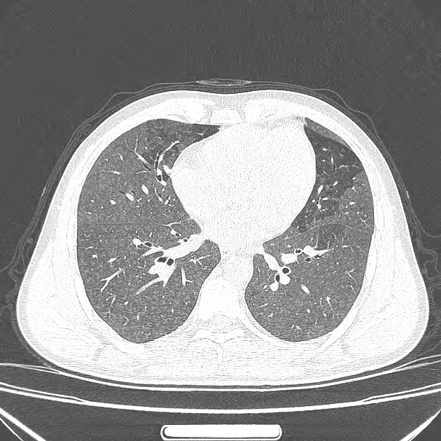
Radiographic features
Plain radiograph
Chest radiographs can be normal in the early stages. There may be ill-defined or ground-glass opacities with lower lobe distribution or consolidation in a patchy, reticulonodular or mixed pattern.
CT
Imaging features can overlap between cellular and fibrotic types, as well as usual interstitial pneumonia (UIP), in as many as 30% of patients. Also, temporal changes in the pattern of HRCT findings in subsequent studies are shown in up to 28% of cases, which result in changes in the provisional diagnosis from NSIP to UIP.
Involvement tends to be subpleural and generally symmetrical with an apicobasal gradient. Solely or predominantly upper lobe involvement or purely unilateral disease makes the diagnosis of NSIP less likely.
Common manifestations include:
- ground-glass opacities
- tends to be a dominant feature: can be symmetrically or diffusely distributed in all zones or display a basal predominance
- immediate subpleural sparing: relatively specific sign
- fine reticular opacities
Following imaging features are more common in fibrotic type:
- thickening of bronchovascular bundles
- traction bronchiectasis: associated with fibrotic NSIP
- lung volume loss: particularly lower lobes
Treatment and prognosis
Cellular type usually responds very well to corticosteroids with ~100% 10-year survival.
Differential diagnosis
-
hypersensitivity pneumonitis
- cellular NSIP can be indistinguishable from
- acute/inflammatory type of hypersensitivity pneumonitis or
- non fibrotic hypersensitivity pneumonitis
- cellular NSIP can be indistinguishable from
Related Radiopaedia articles
Chest
- imaging techniques
-
chest x-ray
-
approach
- adult
- pediatric
- neonatal
-
airspace opacification
- differential diagnoses of airspace opacification
- lobar consolidation
-
atelectasis
- mechanism-based
- morphology-based
- lobar lung collapse
- chest x-ray in the exam setting
- cardiomediastinal contour
- chest radiograph zones
- tracheal air column
- fissures
- normal chest x-ray appearance of the diaphragm
- nipple shadow
-
lines and stripes
- anterior junction line
- posterior junction line
- right paratracheal stripe
- left paratracheal stripe
- posterior tracheal stripe/tracheo-esophageal stripe
- posterior wall of bronchus intermedius
- right paraspinal line
- left paraspinal line
- aortic-pulmonary stripe
- aortopulmonary window
- azygo-esophageal recess
- spaces
- signs
- air bronchogram
- big rib sign
- Chang sign
- Chen sign
- coin lesion
- continuous diaphragm sign
- dense hilum sign
- double contour sign
- egg-on-a-string sign
- extrapleural sign
- finger in glove sign
- flat waist sign
- Fleischner sign
- ginkgo leaf sign
- Golden S sign
- Hampton hump
- haystack sign
- hilum convergence sign
- hilum overlay sign
- Hoffman-Rigler sign
- holly leaf sign
- incomplete border sign
- juxtaphrenic peak sign
- Kirklin sign
- medial stripe sign
- melting ice cube sign
- more black sign
- Naclerio V sign
- Palla sign
- pericardial fat tag sign
- Shmoo sign
- silhouette sign
- snowman sign
- spinnaker sign
- steeple sign
- straight left heart border sign
- third mogul sign
- tram-track sign
- walking man sign
- water bottle sign
- wave sign
- Westermark sign
-
approach
- HRCT
-
chest x-ray
- airways
- bronchitis
- small airways disease
-
bronchiectasis
- broncho-arterial ratio
- related conditions
- differentials by distribution
- narrowing
-
tracheal stenosis
- diffuse tracheal narrowing (differential)
-
bronchial stenosis
- diffuse airway narrowing (differential)
-
tracheal stenosis
- diverticula
- pulmonary edema
-
interstitial lung disease (ILD)
- drug-induced interstitial lung disease
-
hypersensitivity pneumonitis
- acute hypersensitivity pneumonitis
- subacute hypersensitivity pneumonitis
- chronic hypersensitivity pneumonitis
- etiology
- bird fancier's lung: pigeon fancier's lung
- farmer's lung
- cheese workers' lung
- bagassosis
- mushroom worker’s lung
- malt worker’s lung
- maple bark disease
- hot tub lung
- wine maker’s lung
- woodsman’s disease
- thatched roof lung
- tobacco grower’s lung
- potato riddler’s lung
- summer-type pneumonitis
- dry rot lung
- machine operator’s lung
- humidifier lung
- shower curtain disease
- furrier’s lung
- miller’s lung
- lycoperdonosis
- saxophone lung
-
idiopathic interstitial pneumonia (mnemonic)
- acute interstitial pneumonia (AIP)
- cryptogenic organizing pneumonia (COP)
- desquamative interstitial pneumonia (DIP)
- non-specific interstitial pneumonia (NSIP)
- idiopathic pleuroparenchymal fibroelastosis
- lymphoid interstitial pneumonia (LIP)
- respiratory bronchiolitis–associated interstitial lung disease (RB-ILD)
- usual interstitial pneumonia / idiopathic pulmonary fibrosis (UIP/IPF)
-
pneumoconioses
- fibrotic
- non-fibrotic
-
lung cancer
-
non-small-cell lung cancer
-
adenocarcinoma
- pre-invasive tumors
- minimally invasive tumors
- invasive tumors
- variants of invasive carcinoma
- described imaging features
- adenosquamous carcinoma
- large cell carcinoma
- primary sarcomatoid carcinoma of the lung
- squamous cell carcinoma
- salivary gland-type tumors
-
adenocarcinoma
- pulmonary neuroendocrine tumors
- preinvasive lesions
-
lung cancer invasion patterns
- tumor spread through air spaces (STAS)
- presence of non-lepidic patterns such as acinar, papillary, solid, or micropapillary
- myofibroblastic stroma associated with invasive tumor cells
- pleural invasion
- vascular invasion
- tumors by location
- benign neoplasms
- pulmonary metastases
- lung cancer screening
- lung cancer staging
-
non-small-cell lung cancer