
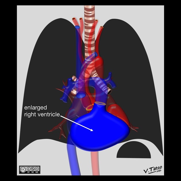
Right ventricular enlargement
Citation, DOI, disclosures and article data
At the time the article was created The Radswiki had no recorded disclosures.
View The Radswiki's current disclosuresAt the time the article was last revised Arlene Campos had no financial relationships to ineligible companies to disclose.
View Arlene Campos's current disclosures- Right ventricular dilatation
- Right ventricular dilatation (RVD)
Right ventricular enlargement (also known as right ventricular dilatation (RVD)) can be the result of a number of conditions, including:
Clinical presentation
ECG
right axis deviation
secondary repolarization (ST-T) abnormalities (leads V1-3)
deep S waves in lateral (I, aVL, V5-6) leads
-
tall, prominent R waves in lead V1
may be masked in the presence of COPD
Radiographic features
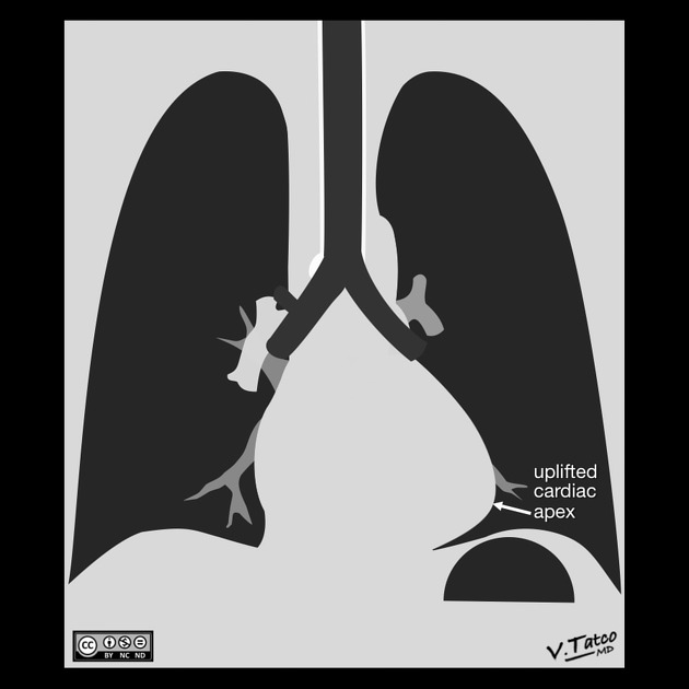
Plain radiograph
Frontal view demonstrates:
rounded left heart border
uplifted cardiac apex
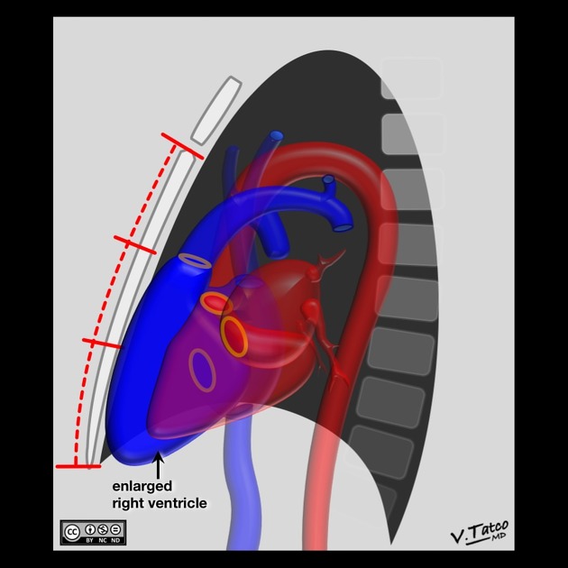
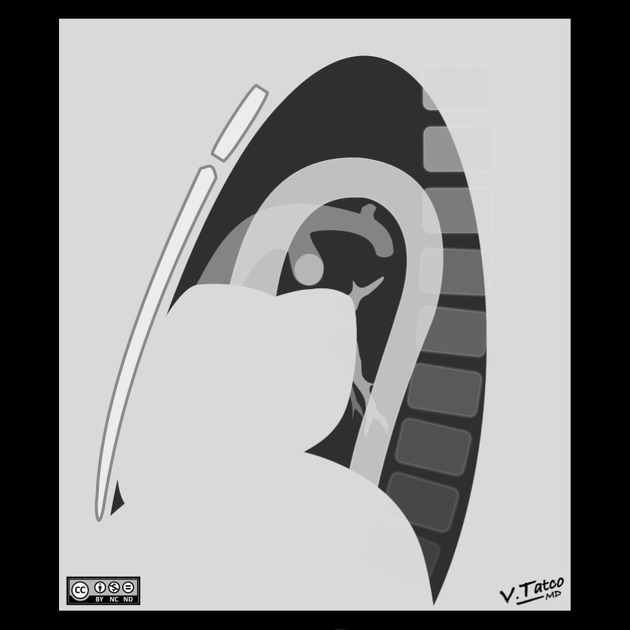
Lateral view demonstrates:
filling of the retrosternal space
rotation of the heart posteriorly
Ultrasound
Echocardiography
On transthoracic echocardiography the apical 4 chamber (A4C) view allows a qualitative assessment of the presence or absence of right ventricular enlargement, as well as the degree of severity 4:
-
mild RV enlargement
basal diameter increased (>4.2 cm)
left ventricular size still exceeds that of the RV
-
moderate RV enlargement
the size of the RV approximates that of the left ventricle (LV)
the left ventricle still forms apex
-
severe RV enlargement
the RV forms the apex and is larger than the LV
References
- 1. Cardiac Imaging: The Requisites. (2004) ISBN: 032301755X - Google Books
- 2. William E. Brant, Clyde A. Helms. Fundamentals of Diagnostic Radiology. (2012) ISBN: 9781608319114 - Google Books
- 3. W. Richard Webb, Charles B. Higgins. Thoracic Imaging. (2011) ISBN: 9781605479767 - Google Books
- 4. Dandel M & Hetzer R. Evaluation of the Right Ventricle by Echocardiography: Particularities and Major Challenges. Expert Rev Cardiovasc Ther. 2018;16(4):259-75. doi:10.1080/14779072.2018.1449646 - Pubmed
Incoming Links
- Pulmonary arterial atherosclerosis
- Right heart strain
- Cardiomegaly
- Total anomalous pulmonary venous return
- Tetralogy of Fallot
- Pulmonary emphysema
- Medical abbreviations and acronyms (R)
- Pulmonary valve stenosis
- Ankylosing spondylitis (cardiovascular manifestations)
- Uhl anomaly
- Cardiac blood pool scan
- Chronic pulmonary embolism
- Pulmonary valve regurgitation
- Double outlet right ventricle
- Tricuspid valve regurgitation
- Cardiac chamber enlargement
Related articles: Chest
- imaging techniques
-
chest radiograph
- radiography[+][+]
-
approach
- ABCDE
- ABCDEFGHI
- congenital heart disease
- medical devices in the thorax
- common lines and tubes[+][+]
- nasogastric tubes
- endotracheal tubes
- central venous catheters
- esophageal temperature probe
- tracheostomy tube
- pleural catheters
- cardiac conduction devices
- prosthetic heart valve
- review areas
-
airspace opacification[+][+]
- differential diagnoses of airspace opacification
- lobar consolidation
-
atelectasis
- mechanism-based
- morphology-based
- lobar lung collapse
- chest x-ray in the exam setting[+][+]
- cardiomediastinal contour
- chest radiograph zones[+][+]
- tracheal air column[+][+]
- fissures[+][+]
- normal chest x-ray appearance of the diaphragm[+][+]
- nipple shadow[+][+]
-
lines and stripes[+][+]
- anterior junction line
- posterior junction line
- right paratracheal stripe
- left paratracheal stripe
- posterior tracheal stripe/tracheo-esophageal stripe
- posterior wall of bronchus intermedius
- right paraspinal line
- left paraspinal line
- aortic-pulmonary stripe
- aortopulmonary window
- azygo-esophageal recess
- spaces[+][+]
- signs[+][+]
- air bronchogram
- big rib sign
- Chang sign
- Chen sign
- coin lesion
- continuous diaphragm sign
- dense hilum sign
- double contour sign
- egg-on-a-string sign
- extrapleural sign
- finger in glove sign
- flat waist sign
- Fleischner sign
- ginkgo leaf sign
- Golden S sign
- Hampton hump
- haystack sign
- hilum convergence sign
- hilum overlay sign
- Hoffman-Rigler sign
- holly leaf sign
- incomplete border sign
- juxtaphrenic peak sign
- Kirklin sign
- medial stripe sign
- melting ice cube sign
- more black sign
- Naclerio V sign
- Palla sign
- pericardial fat tag sign
- Shmoo sign
- silhouette sign
- snowman sign
- spinnaker sign
- steeple sign
- straight left heart border sign
- third mogul sign
- tram-track sign
- walking man sign
- water bottle sign
- wave sign
- Westermark sign
- HRCT[+][+]
-
chest radiograph
- airways[+][+]
- bronchitis
- small airways disease
-
bronchiectasis
- broncho-arterial ratio
- related conditions
- differentials by distribution
- narrowing
-
tracheal stenosis
- diffuse tracheal narrowing (differential)
-
bronchial stenosis
- diffuse airway narrowing (differential)
-
tracheal stenosis
- diverticula
- pulmonary edema[+][+]
-
interstitial lung disease (ILD)[+][+]
- Anti-Jo-1 antibody-positive interstitial lung disease
- drug-induced interstitial lung disease
-
hypersensitivity pneumonitis
- acute hypersensitivity pneumonitis
- subacute hypersensitivity pneumonitis
- chronic hypersensitivity pneumonitis
- etiology
- bird fancier's lung: pigeon fancier's lung
- farmer's lung
- cheese workers' lung
- bagassosis
- mushroom worker’s lung
- malt worker’s lung
- maple bark disease
- hot tub lung
- wine maker’s lung
- woodsman’s disease
- thatched roof lung
- tobacco grower’s lung
- potato riddler’s lung
- summer-type pneumonitis
- dry rot lung
- machine operator’s lung
- humidifier lung
- shower curtain disease
- furrier’s lung
- miller’s lung
- lycoperdonosis
- saxophone lung
-
idiopathic interstitial pneumonia (mnemonic)
- acute interstitial pneumonia (AIP)
- cryptogenic organizing pneumonia (COP)
- desquamative interstitial pneumonia (DIP)
- non-specific interstitial pneumonia (NSIP)
- idiopathic pleuroparenchymal fibroelastosis
- lymphoid interstitial pneumonia (LIP)
- respiratory bronchiolitis–associated interstitial lung disease (RB-ILD)
- usual interstitial pneumonia / idiopathic pulmonary fibrosis (UIP/IPF)
-
pneumoconioses
- fibrotic
- non-fibrotic
-
lung cancer[+][+]
-
non-small-cell lung cancer
-
adenocarcinoma
- pre-invasive tumors
- minimally invasive tumors
- invasive tumors
- variants of invasive carcinoma
- described imaging features
- adenosquamous carcinoma
- large cell carcinoma
- primary sarcomatoid carcinoma of the lung
- squamous cell carcinoma
- salivary gland-type tumors
-
adenocarcinoma
- pulmonary neuroendocrine tumors
- preinvasive lesions
-
lung cancer invasion patterns
- tumor spread through air spaces (STAS)
- presence of non-lepidic patterns such as acinar, papillary, solid, or micropapillary
- myofibroblastic stroma associated with invasive tumor cells
- pleural invasion
- vascular invasion
- tumors by location
- benign neoplasms
- pulmonary metastases
- lung cancer screening
- lung cancer staging
-
non-small-cell lung cancer


 Unable to process the form. Check for errors and try again.