
Gravity-dependent atelectasis
Citation, DOI, disclosures and article data
At the time the article was created Craig Hacking had no recorded disclosures.
View Craig Hacking's current disclosuresAt the time the article was last revised Henry Knipe had the following disclosures:
- Radiopaedia Events Pty Ltd, Speaker fees (past)
- Integral Diagnostics, Shareholder (ongoing)
- Micro-X Ltd, Shareholder (ongoing)
These were assessed during peer review and were determined to not be relevant to the changes that were made.
View Henry Knipe's current disclosures- Gravity dependent atelectasis
- Dependant lung atelectasis
- Dependent lung atelectasis
- Gravity dependent atelectasis
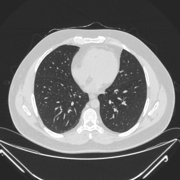
Gravity-dependent atelectasis refers to a form of lung atelectasis that occurs in the dependent portions of the lungs.
On this page:
Images:
Pathology
Gravity-dependent atelectasis occurs due to a combination of reduced alveolar volume and increased perfusion. Due to gravity, it usually has a dependent and subpleural distribution. It is very commonly seen in the posterior lung bases on CT, particularly in elderly individuals.
In normal lung, gravity gradients exist in end-inspiration between the apex and lung base of 4:1 in the erect patient and between the anterior and posterior lung of 2.5:1 in the supine patient 1. These gradients increase in the presence of lung disease that increases the weight of the lung causing atelectasis.
Pathology
Etiology
bedridden patients
patients with prolonged shallow breathing
impaired mucociliary clearance
Differential diagnosis
early interstitial lung disease: prone CT chest can usually differentiate 2
References
- 1. Woodring J & Reed J. Types and Mechanisms of Pulmonary Atelectasis. J Thorac Imaging. 1996;11(2):92-108. doi:10.1097/00005382-199621000-00002 - Pubmed
- 2. Kashiwabara K & Kohshi S. Additional Computed Tomography Scans in the Prone Position to Distinguish Early Interstitial Lung Disease from Dependent Density on Helical Computed Tomography Screening Patient Characteristics. Respirology. 2006;11(4):482-7. doi:10.1111/j.1440-1843.2006.00869.x - Pubmed
Incoming Links
Related articles: Airspace opacification
- airspace opacification
- differential diagnoses of airspace opacification[+][+]
- lobar consolidation[+][+]
-
atelectasis
-
mechanism-based
- resorptive (obstructive) atelectasis
- passive (relaxation) atelectasis
- compressive atelectasis
- cicatrisation atelectasis
- adhesive atelectasis
- gravity dependant atelectasis
- morphology-based[+][+]
-
mechanism-based
- lobar lung collapse[+][+]
Related articles: Chest
- imaging techniques
-
chest radiograph
- radiography[+][+]
-
approach
- ABCDE
- ABCDEFGHI
- congenital heart disease
- medical devices in the thorax
- common lines and tubes[+][+]
- nasogastric tubes
- endotracheal tubes
- central venous catheters
- esophageal temperature probe
- tracheostomy tube
- pleural catheters
- cardiac conduction devices
- prosthetic heart valve
- review areas
-
airspace opacification
- differential diagnoses of airspace opacification[+][+]
- lobar consolidation[+][+]
-
atelectasis
-
mechanism-based
- resorptive (obstructive) atelectasis
- passive (relaxation) atelectasis
- compressive atelectasis
- cicatrisation atelectasis
- adhesive atelectasis
- gravity dependant atelectasis
- morphology-based[+][+]
-
mechanism-based
- lobar lung collapse[+][+]
- chest x-ray in the exam setting[+][+]
- cardiomediastinal contour[+][+]
- chest radiograph zones[+][+]
- tracheal air column[+][+]
- fissures[+][+]
- normal chest x-ray appearance of the diaphragm[+][+]
- nipple shadow[+][+]
-
lines and stripes[+][+]
- anterior junction line
- posterior junction line
- right paratracheal stripe
- left paratracheal stripe
- posterior tracheal stripe/tracheo-esophageal stripe
- posterior wall of bronchus intermedius
- right paraspinal line
- left paraspinal line
- aortic-pulmonary stripe
- aortopulmonary window
- azygo-esophageal recess
- spaces[+][+]
- signs[+][+]
- air bronchogram
- big rib sign
- Chang sign
- Chen sign
- coin lesion
- continuous diaphragm sign
- dense hilum sign
- double contour sign
- egg-on-a-string sign
- extrapleural sign
- finger in glove sign
- flat waist sign
- Fleischner sign
- ginkgo leaf sign
- Golden S sign
- Hampton hump
- haystack sign
- hilum convergence sign
- hilum overlay sign
- Hoffman-Rigler sign
- holly leaf sign
- incomplete border sign
- juxtaphrenic peak sign
- Kirklin sign
- medial stripe sign
- melting ice cube sign
- more black sign
- Naclerio V sign
- Palla sign
- pericardial fat tag sign
- Shmoo sign
- silhouette sign
- snowman sign
- spinnaker sign
- steeple sign
- straight left heart border sign
- third mogul sign
- tram-track sign
- walking man sign
- water bottle sign
- wave sign
- Westermark sign
- HRCT[+][+]
-
chest radiograph
- airways[+][+]
- bronchitis
- small airways disease
-
bronchiectasis
- broncho-arterial ratio
- related conditions
- differentials by distribution
- narrowing
-
tracheal stenosis
- diffuse tracheal narrowing (differential)
-
bronchial stenosis
- diffuse airway narrowing (differential)
-
tracheal stenosis
- diverticula
- pulmonary edema[+][+]
-
interstitial lung disease (ILD)[+][+]
- Anti-Jo-1 antibody-positive interstitial lung disease
- drug-induced interstitial lung disease
-
hypersensitivity pneumonitis
- acute hypersensitivity pneumonitis
- subacute hypersensitivity pneumonitis
- chronic hypersensitivity pneumonitis
- etiology
- bird fancier's lung: pigeon fancier's lung
- farmer's lung
- cheese workers' lung
- bagassosis
- mushroom worker’s lung
- malt worker’s lung
- maple bark disease
- hot tub lung
- wine maker’s lung
- woodsman’s disease
- thatched roof lung
- tobacco grower’s lung
- potato riddler’s lung
- summer-type pneumonitis
- dry rot lung
- machine operator’s lung
- humidifier lung
- shower curtain disease
- furrier’s lung
- miller’s lung
- lycoperdonosis
- saxophone lung
-
idiopathic interstitial pneumonia (mnemonic)
- acute interstitial pneumonia (AIP)
- cryptogenic organizing pneumonia (COP)
- desquamative interstitial pneumonia (DIP)
- non-specific interstitial pneumonia (NSIP)
- idiopathic pleuroparenchymal fibroelastosis
- lymphoid interstitial pneumonia (LIP)
- respiratory bronchiolitis–associated interstitial lung disease (RB-ILD)
- usual interstitial pneumonia / idiopathic pulmonary fibrosis (UIP/IPF)
-
pneumoconioses
- fibrotic
- non-fibrotic
-
lung cancer[+][+]
-
non-small-cell lung cancer
-
adenocarcinoma
- pre-invasive tumors
- minimally invasive tumors
- invasive tumors
- variants of invasive carcinoma
- described imaging features
- adenosquamous carcinoma
- large cell carcinoma
- primary sarcomatoid carcinoma of the lung
- squamous cell carcinoma
- salivary gland-type tumors
-
adenocarcinoma
- pulmonary neuroendocrine tumors
- preinvasive lesions
-
lung cancer invasion patterns
- tumor spread through air spaces (STAS)
- presence of non-lepidic patterns such as acinar, papillary, solid, or micropapillary
- myofibroblastic stroma associated with invasive tumor cells
- pleural invasion
- vascular invasion
- tumors by location
- benign neoplasms
- pulmonary metastases
- lung cancer screening
- lung cancer staging
-
non-small-cell lung cancer


 Unable to process the form. Check for errors and try again.