Cryptogenic organizing pneumonia
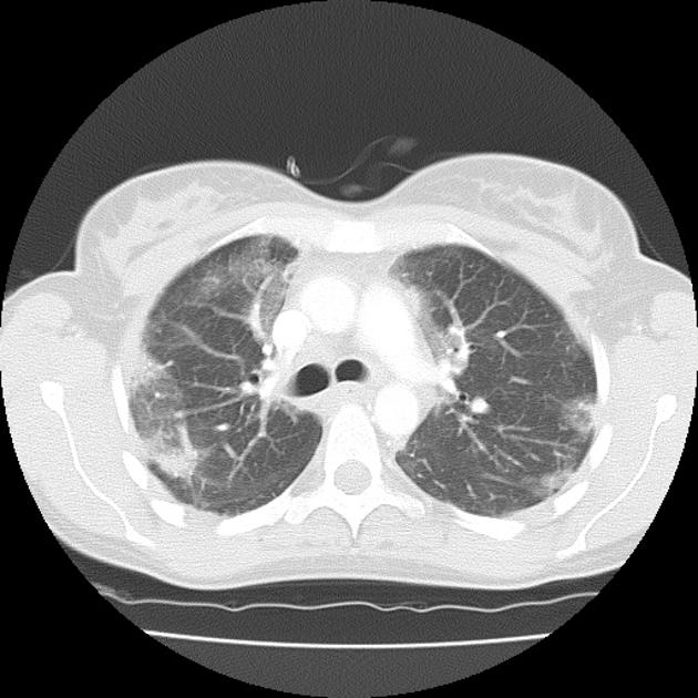
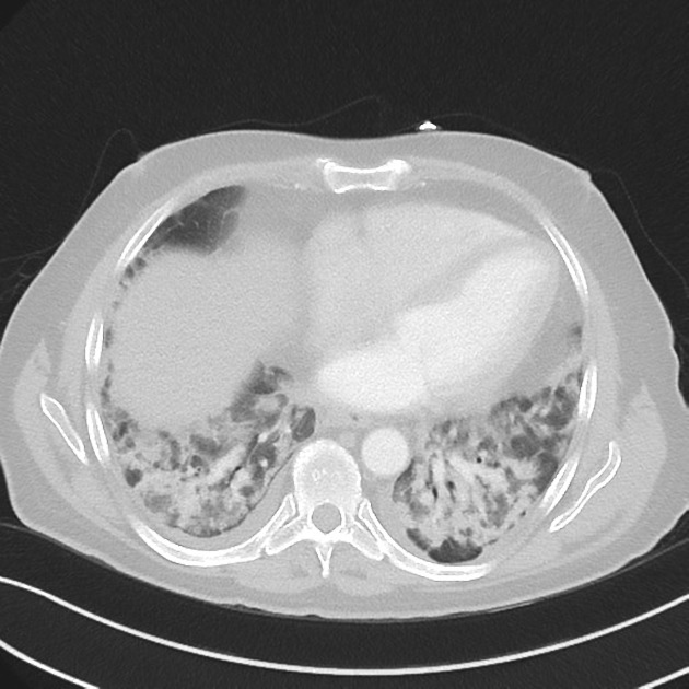
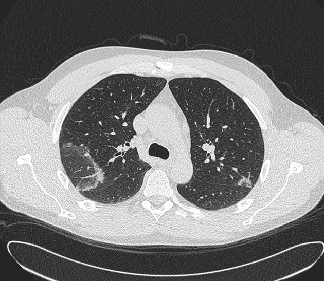
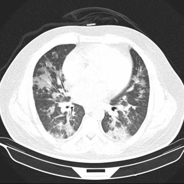
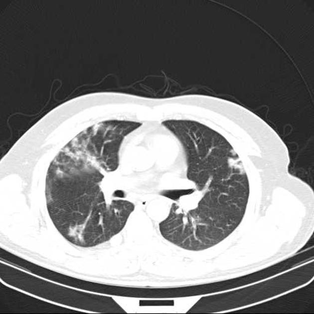
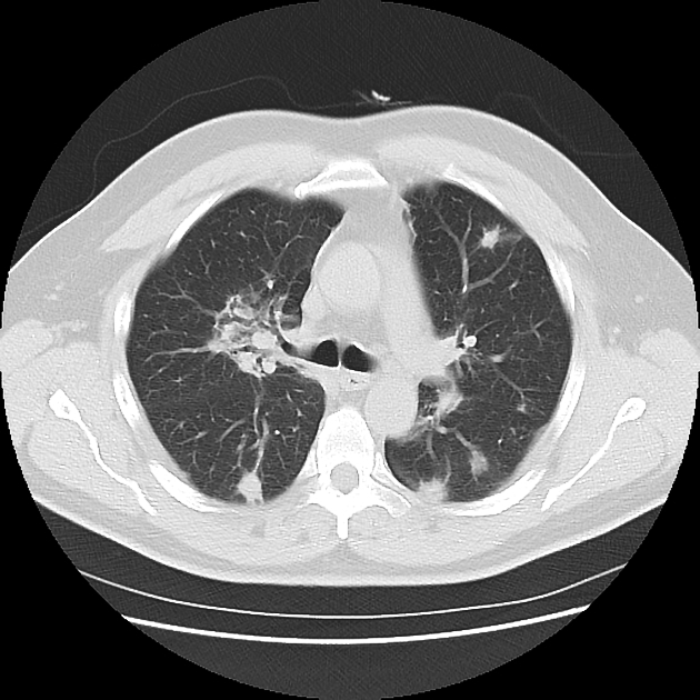
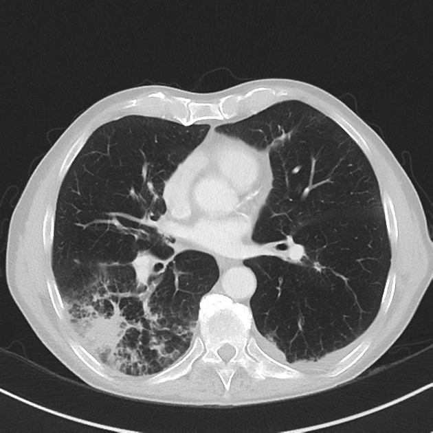
Cryptogenic organizing pneumonia (COP) is a disease of unknown etiology characterized on imaging by multifocal ground glass opacifications and/or consolidation. A wide variety of infectious as well as noninfectious causes may result in a similar histologic pattern.
On this page:
Terminology
Organizing pneumonia (OP) is a histological pattern of alveolar inflammation with varied etiology (including pulmonary infection). The idiopathic form of OP is called cryptogenic organizing pneumonia (COP) and it belongs to the idiopathic interstitial pneumonias (IIPs).
COP was previously termed bronchiolitis obliterans organizing pneumonia (BOOP), not to be confused with bronchiolitis obliterans 8.
Epidemiology
The presentation is commonest in the 55-60 years age group.
Clinical presentation
Patients present with a short history (i.e. less than ~2 months) of breathlessness, non-productive cough, weight loss, malaise and fever. There is no association with smoking.
Pathology
In addition to the alveolar inflammatory changes found with regular pneumonia, there is also the involvement of the bronchioles.
Histologically, it is characterized by mild chronic patchy interstitial inflammation without fibrosis and the presence of buds of granulation tissue made of mononuclear cells, foamy macrophages, and fibrous tissue (Masson bodies) in the distal airspaces which may cause secondary bronchiolar occlusion due to extension of the inflammatory process. Hence, the reason for being previously termed bronchiolitis obliterans organizing pneumonia (BOOP).
Radiographic features
Plain radiograph
- consolidation
- unilateral or bilateral patchy areas (commonest finding 3): often migratory
- can affect all lung zones
- usually peripheral, subpleural, peribronchovascular 2
- nodules
- foci of granulation tissue up to 1 cm
- may mimic neoplasm if >5 cm in size
- may be numerous in immunocompromised patients
CT
The most common HRCT features include 6:
- patchy consolidation with a predominantly subpleural and/or peribronchial distribution
- small, ill-defined peribronchial or peribronchiolar nodules
- large nodules or masses
- bronchial wall thickening or dilatation in the abnormal lung regions
- a perilobular pattern with ill-defined linear opacities that are thicker than the thickened interlobular septa and have an arcade or polygonal appearance
- arcade-like sign of perilobular fibrosis describes an arch pattern in more than half of the patient with COP 13
- ground glass opacity or crazy paving
The reverse halo sign (atoll sign) is considered to be highly specific, although only seen in ~20% of patients with COP 5.
Treatment and prognosis
Corticosteroids have been widely used and most patients recover completely 3-4.
History and etymology
It was first described by Davison and colleagues in 1983.
Differential diagnosis
On a radiograph consider:
- differential for peripheral consolidation: reverse bat wing opacities
- differential for bilateral airspace opacities
On CT consider:
- adenocarcinoma in situ or minimally invasive (formerly bronchoalveolar carcinoma)
- pulmonary lymphoma
- pulmonary vasculitis/vasculitides
- sarcoidosis
- chronic eosinophilic pneumonia (for a subpleural consolidative pattern)
Related Radiopaedia articles
Chest
- imaging techniques
-
chest x-ray
-
approach
- adult
- pediatric
- neonatal
-
airspace opacification
- differential diagnoses of airspace opacification
- lobar consolidation
-
atelectasis
- mechanism-based
- morphology-based
- lobar lung collapse
- chest x-ray in the exam setting
- cardiomediastinal contour
- chest radiograph zones
- tracheal air column
- fissures
- normal chest x-ray appearance of the diaphragm
- nipple shadow
-
lines and stripes
- anterior junction line
- posterior junction line
- right paratracheal stripe
- left paratracheal stripe
- posterior tracheal stripe/tracheo-esophageal stripe
- posterior wall of bronchus intermedius
- right paraspinal line
- left paraspinal line
- aortic-pulmonary stripe
- aortopulmonary window
- azygo-esophageal recess
- spaces
- signs
- air bronchogram
- big rib sign
- Chang sign
- Chen sign
- coin lesion
- continuous diaphragm sign
- dense hilum sign
- double contour sign
- egg-on-a-string sign
- extrapleural sign
- finger in glove sign
- flat waist sign
- Fleischner sign
- ginkgo leaf sign
- Golden S sign
- Hampton hump
- haystack sign
- hilum convergence sign
- hilum overlay sign
- Hoffman-Rigler sign
- holly leaf sign
- incomplete border sign
- juxtaphrenic peak sign
- Kirklin sign
- medial stripe sign
- melting ice cube sign
- more black sign
- Naclerio V sign
- Palla sign
- pericardial fat tag sign
- Shmoo sign
- silhouette sign
- snowman sign
- spinnaker sign
- steeple sign
- straight left heart border sign
- third mogul sign
- tram-track sign
- walking man sign
- water bottle sign
- wave sign
- Westermark sign
-
approach
- HRCT
-
chest x-ray
- airways
- bronchitis
- small airways disease
-
bronchiectasis
- broncho-arterial ratio
- related conditions
- differentials by distribution
- narrowing
-
tracheal stenosis
- diffuse tracheal narrowing (differential)
-
bronchial stenosis
- diffuse airway narrowing (differential)
-
tracheal stenosis
- diverticula
- pulmonary edema
-
interstitial lung disease (ILD)
- drug-induced interstitial lung disease
-
hypersensitivity pneumonitis
- acute hypersensitivity pneumonitis
- subacute hypersensitivity pneumonitis
- chronic hypersensitivity pneumonitis
- etiology
- bird fancier's lung: pigeon fancier's lung
- farmer's lung
- cheese workers' lung
- bagassosis
- mushroom worker’s lung
- malt worker’s lung
- maple bark disease
- hot tub lung
- wine maker’s lung
- woodsman’s disease
- thatched roof lung
- tobacco grower’s lung
- potato riddler’s lung
- summer-type pneumonitis
- dry rot lung
- machine operator’s lung
- humidifier lung
- shower curtain disease
- furrier’s lung
- miller’s lung
- lycoperdonosis
- saxophone lung
-
idiopathic interstitial pneumonia (mnemonic)
- acute interstitial pneumonia (AIP)
- cryptogenic organizing pneumonia (COP)
- desquamative interstitial pneumonia (DIP)
- non-specific interstitial pneumonia (NSIP)
- idiopathic pleuroparenchymal fibroelastosis
- lymphoid interstitial pneumonia (LIP)
- respiratory bronchiolitis–associated interstitial lung disease (RB-ILD)
- usual interstitial pneumonia / idiopathic pulmonary fibrosis (UIP/IPF)
-
pneumoconioses
- fibrotic
- non-fibrotic
-
lung cancer
-
non-small-cell lung cancer
-
adenocarcinoma
- pre-invasive tumors
- minimally invasive tumors
- invasive tumors
- variants of invasive carcinoma
- described imaging features
- adenosquamous carcinoma
- large cell carcinoma
- primary sarcomatoid carcinoma of the lung
- squamous cell carcinoma
- salivary gland-type tumors
-
adenocarcinoma
- pulmonary neuroendocrine tumors
- preinvasive lesions
-
lung cancer invasion patterns
- tumor spread through air spaces (STAS)
- presence of non-lepidic patterns such as acinar, papillary, solid, or micropapillary
- myofibroblastic stroma associated with invasive tumor cells
- pleural invasion
- vascular invasion
- tumors by location
- benign neoplasms
- pulmonary metastases
- lung cancer screening
- lung cancer staging
-
non-small-cell lung cancer