
Left ventricular enlargement
Citation, DOI, disclosures and article data
At the time the article was created Frank Gaillard had no recorded disclosures.
View Frank Gaillard's current disclosuresAt the time the article was last revised Yuranga Weerakkody had no recorded disclosures.
View Yuranga Weerakkody's current disclosures- LV enlargement
- LV dilatation
Left ventricular enlargement can be the result of a number of conditions, including:
- pressure overload
- volume overload
- wall abnormalities
Radiographic features
Plain radiograph
Features that may be visible on a chest radiograph include:
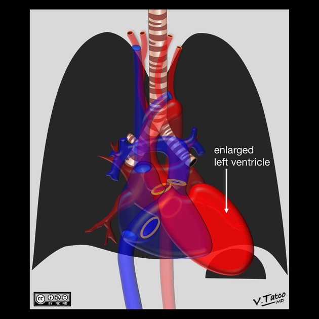
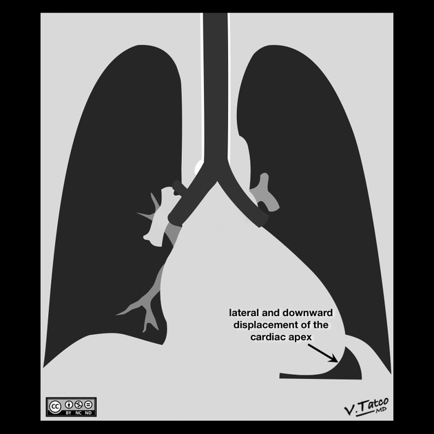
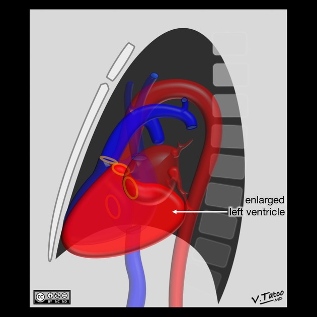
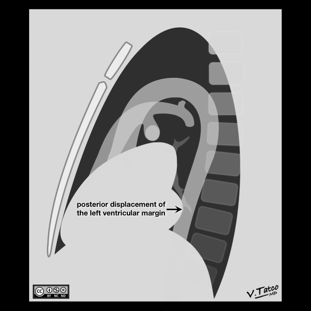
- left ventricular dilatation: left heart border is displaced leftward, inferiorly and posteriorly
- left ventricular hypertrophy: may show rounding of the cardiac apex
- Hoffman-Rigler sign
- Shmoo sign
Echocardiography
The parasternal long axis and apical four-chamber views on transthoracic echocardiography are often the primary views used to gain both a qualitative and quantitative appreciation of left ventricular enlargement.
Features include 4:
- increased left ventricular internal end-diastolic diameter (LVIDd)
- parasternal long axis LVIDd >5.3 cm (females) or >5.9 cm (males)
- elevated left ventricular volumes
- diastolic volumes >104 mL (females) or >155 mL (males)
- systolic volumes >49 mL (females) or >58 mL (males)
- increasingly spherical morphology
- a normal left ventricle has prolate ellipsoidal morphology, with a long axis roughly twice that of the short axis
- with severe LV enlargement the short axis dimensions may approximate those of the long axis, akin to a sphere
CT
One publication has suggested left ventricular enlargement being able to be reliably identified on non-gated contrast-enhanced multidetector CT (with sensitivity of 78% and specificity of 100%) when the maximum luminal diameter of the LV is greater than 5.6 cm 5.
References
- 1. Miller SW. Cardiac imaging, the requisites. Mosby Inc. (2005) ISBN:032301755X. Read it at Google Books - Find it at Amazon
- 2. Brant WE, Helms C. Fundamentals of Diagnostic Radiology. LWW. ISBN:1608319113. Read it at Google Books - Find it at Amazon
- 3. Webb WR, Higgins CB. Thoracic Imaging. Lippincott Williams & Wilkins. (2010) ISBN:1605479764. Read it at Google Books - Find it at Amazon
- 4. Kou S, Caballero L, Dulgheru R, Voilliot D, De Sousa C, Kacharava G, Athanassopoulos GD, Barone D, Baroni M, Cardim N, Gomez De Diego JJ, Hagendorff A, Henri C, Hristova K, Lopez T, Magne J, De La Morena G, Popescu BA, Penicka M, Ozyigit T, Rodrigo Carbonero JD, Salustri A, Van De Veire N, Von Bardeleben RS, Vinereanu D, Voigt JU, Zamorano JL, Donal E, Lang RM, Badano LP, Lancellotti P. Echocardiographic reference ranges for normal cardiac chamber size: results from the NORRE study. (2014) European heart journal cardiovascular Imaging. 15 (6): 680-90. doi:10.1093/ehjci/jet284 - Pubmed
- 5. Kathiria N, Devcic Z, Chen J et al. Assessment of Left Ventricular Enlargement at Multidetector Computed Tomography. J Comput Assist Tomogr. 2015;39(5):794-6. doi:10.1097/RCT.0000000000000279 Copy Citation
Incoming Links
- Anomalous left coronary artery from the pulmonary artery
- Hoffman-Rigler sign (heart)
- Cardiac chamber enlargement
- Shmoo sign
- Mitral annular dilation
- Mitral valve regurgitation
- Animal and animal produce inspired signs
- Cardiomegaly
- Cardiac silhouette
- Barth syndrome
- Left ventricle
- Acute coronary syndrome
Related articles: Chest
- imaging techniques
-
chest radiograph
- radiography[+][+]
-
approach
- ABCDE
- ABCDEFGHI
- congenital heart disease
- medical devices in the thorax
- common lines and tubes[+][+]
- nasogastric tubes
- endotracheal tubes
- central venous catheters
- esophageal temperature probe
- tracheostomy tube
- pleural catheters
- cardiac conduction devices
- prosthetic heart valve
- review areas
-
airspace opacification[+][+]
- differential diagnoses of airspace opacification
- lobar consolidation
-
atelectasis
- mechanism-based
- morphology-based
- lobar lung collapse
- chest x-ray in the exam setting[+][+]
- cardiomediastinal contour
- chest radiograph zones[+][+]
- tracheal air column[+][+]
- fissures[+][+]
- normal chest x-ray appearance of the diaphragm[+][+]
- nipple shadow[+][+]
-
lines and stripes[+][+]
- anterior junction line
- posterior junction line
- right paratracheal stripe
- left paratracheal stripe
- posterior tracheal stripe/tracheo-esophageal stripe
- posterior wall of bronchus intermedius
- right paraspinal line
- left paraspinal line
- aortic-pulmonary stripe
- aortopulmonary window
- azygo-esophageal recess
- spaces[+][+]
- signs[+][+]
- air bronchogram
- big rib sign
- Chang sign
- Chen sign
- coin lesion
- continuous diaphragm sign
- dense hilum sign
- double contour sign
- egg-on-a-string sign
- extrapleural sign
- finger in glove sign
- flat waist sign
- Fleischner sign
- ginkgo leaf sign
- Golden S sign
- Hampton hump
- haystack sign
- hilum convergence sign
- hilum overlay sign
- Hoffman-Rigler sign
- holly leaf sign
- incomplete border sign
- juxtaphrenic peak sign
- Kirklin sign
- medial stripe sign
- melting ice cube sign
- more black sign
- Naclerio V sign
- Palla sign
- pericardial fat tag sign
- Shmoo sign
- silhouette sign
- snowman sign
- spinnaker sign
- steeple sign
- straight left heart border sign
- third mogul sign
- tram-track sign
- walking man sign
- water bottle sign
- wave sign
- Westermark sign
- HRCT[+][+]
-
chest radiograph
- airways[+][+]
- bronchitis
- small airways disease
-
bronchiectasis
- broncho-arterial ratio
- related conditions
- differentials by distribution
- narrowing
-
tracheal stenosis
- diffuse tracheal narrowing (differential)
-
bronchial stenosis
- diffuse airway narrowing (differential)
-
tracheal stenosis
- diverticula
- pulmonary edema[+][+]
-
interstitial lung disease (ILD)[+][+]
- Anti-Jo-1 antibody-positive interstitial lung disease
- drug-induced interstitial lung disease
-
hypersensitivity pneumonitis
- acute hypersensitivity pneumonitis
- subacute hypersensitivity pneumonitis
- chronic hypersensitivity pneumonitis
- etiology
- bird fancier's lung: pigeon fancier's lung
- farmer's lung
- cheese workers' lung
- bagassosis
- mushroom worker’s lung
- malt worker’s lung
- maple bark disease
- hot tub lung
- wine maker’s lung
- woodsman’s disease
- thatched roof lung
- tobacco grower’s lung
- potato riddler’s lung
- summer-type pneumonitis
- dry rot lung
- machine operator’s lung
- humidifier lung
- shower curtain disease
- furrier’s lung
- miller’s lung
- lycoperdonosis
- saxophone lung
-
idiopathic interstitial pneumonia (mnemonic)
- acute interstitial pneumonia (AIP)
- cryptogenic organizing pneumonia (COP)
- desquamative interstitial pneumonia (DIP)
- non-specific interstitial pneumonia (NSIP)
- idiopathic pleuroparenchymal fibroelastosis
- lymphoid interstitial pneumonia (LIP)
- respiratory bronchiolitis–associated interstitial lung disease (RB-ILD)
- usual interstitial pneumonia / idiopathic pulmonary fibrosis (UIP/IPF)
-
pneumoconioses
- fibrotic
- non-fibrotic
-
lung cancer[+][+]
-
non-small-cell lung cancer
-
adenocarcinoma
- pre-invasive tumors
- minimally invasive tumors
- invasive tumors
- variants of invasive carcinoma
- described imaging features
- adenosquamous carcinoma
- large cell carcinoma
- primary sarcomatoid carcinoma of the lung
- squamous cell carcinoma
- salivary gland-type tumors
-
adenocarcinoma
- pulmonary neuroendocrine tumors
- preinvasive lesions
-
lung cancer invasion patterns
- tumor spread through air spaces (STAS)
- presence of non-lepidic patterns such as acinar, papillary, solid, or micropapillary
- myofibroblastic stroma associated with invasive tumor cells
- pleural invasion
- vascular invasion
- tumors by location
- benign neoplasms
- pulmonary metastases
- lung cancer screening
- lung cancer staging
-
non-small-cell lung cancer


 Unable to process the form. Check for errors and try again.