
Review areas on chest radiograph
Citation, DOI, disclosures and article data
At the time the article was created Appukutty Manickam had no recorded disclosures.
View Appukutty Manickam's current disclosuresAt the time the article was last revised Joshua Yap had no financial relationships to ineligible companies to disclose.
View Joshua Yap's current disclosures- Review areas on a chest x-ray
- Review areas on a CXR
- Review areas on chest x-rays
- Chest radiograph review area
- Chest radiograph review areas
- CXR review area
- CXR review areas
- Chest x-ray review areas
- Chest x-ray review area
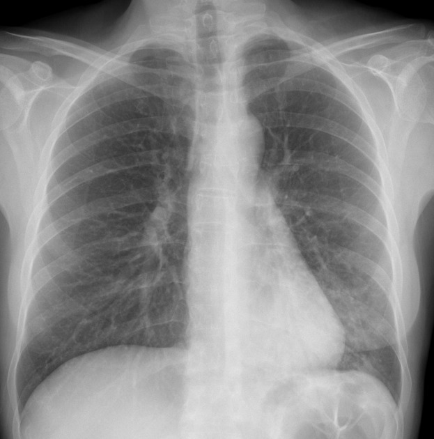
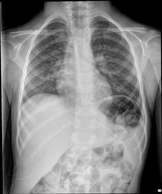
Review areas on a chest radiograph are common areas for missed findings, and special attention should be paid to them:
lung apices: masses (e.g. Pancoast tumor), pneumothorax
behind the heart: consolidation, masses, hiatus hernia 2
below the diaphragm: free gas, lines and tubes (e.g. nasogastric tube), gastric distension, bowel obstruction
bones and soft tissues: fractures, masses, mastectomy, subcutaneous emphysema, evidence of previous surgery (e.g. axillary clips)
These first four are the classic review areas ref, sometimes the pulmonary hilum is included as an additional fifth region:
pulmonary hilum: mass, lymphadenopathy, vessel enlargement
See also
Quiz questions
References
- 1. Elizabeth Puddy, Catherine Hill; Interpretation of the chest radiograph, Continuing Education in Anaesthesia Critical Care & Pain, Volume 7, Issue 3, 1 June 2007, Pages 71–75, https://doi.org/10.1093/bjaceaccp/mkm014
- 2. Oestreich AE. A helpful hint for chest radiology: "look behind the heart". (1982) Journal of the National Medical Association. 74 (10): 1029-31. Pubmed
Incoming Links
- Review area
- Assessment of bones and soft tissue on chest x-ray
- Lines and tubes (radiograph)
- Assessment of pulmonary hila on chest x-ray (approach)
- Assessment of cardiomediastinal contours on chest x-ray (approach)
- Apical zone
- Adult chest radiograph pathology checklist
- Systematic chest radiograph assessment (approach)
- Assessment of chest x-ray technical adequacy (approach)
- Apical chest mass
- Assessment of lungs, pleura and airways on chest x-ray (approach)
- Difficult-to-detect pulmonary nodule
- Mastectomy
- Normal chest radiograph
- Normal chest radiograph
- Normal chest radiograph
- Achalasia
- Underinspiration and poor positioning mimicking lung pathology in a pediatric patient
- T1b apical lung cancer
- Dense hilum sign
- Left apical lung cancer
- Shoulder dislocation (trauma chest x-ray)
- Lung cancer - retrocardiac
- Left lower lobe pneumonia - retrocardiac consolidation
Related articles: Chest
- imaging techniques
-
chest radiograph
- radiography[+][+]
-
approach
- ABCDE
- ABCDEFGHI
- congenital heart disease
- medical devices in the thorax
- common lines and tubes
- nasogastric tubes[+][+]
- endotracheal tubes[+][+]
- central venous catheters[+][+]
- esophageal temperature probe
- tracheostomy tube
- pleural catheters
- cardiac conduction devices
- prosthetic heart valve
- review areas
-
airspace opacification[+][+]
- differential diagnoses of airspace opacification
- lobar consolidation
-
atelectasis
- mechanism-based
- morphology-based
- lobar lung collapse
- chest x-ray in the exam setting[+][+]
- cardiomediastinal contour[+][+]
- chest radiograph zones[+][+]
- tracheal air column[+][+]
- fissures[+][+]
- normal chest x-ray appearance of the diaphragm[+][+]
- nipple shadow[+][+]
-
lines and stripes[+][+]
- anterior junction line
- posterior junction line
- right paratracheal stripe
- left paratracheal stripe
- posterior tracheal stripe/tracheo-esophageal stripe
- posterior wall of bronchus intermedius
- right paraspinal line
- left paraspinal line
- aortic-pulmonary stripe
- aortopulmonary window
- azygo-esophageal recess
- spaces[+][+]
- signs[+][+]
- air bronchogram
- big rib sign
- Chang sign
- Chen sign
- coin lesion
- continuous diaphragm sign
- dense hilum sign
- double contour sign
- egg-on-a-string sign
- extrapleural sign
- finger in glove sign
- flat waist sign
- Fleischner sign
- ginkgo leaf sign
- Golden S sign
- Hampton hump
- haystack sign
- hilum convergence sign
- hilum overlay sign
- Hoffman-Rigler sign
- holly leaf sign
- incomplete border sign
- juxtaphrenic peak sign
- Kirklin sign
- medial stripe sign
- melting ice cube sign
- more black sign
- Naclerio V sign
- Palla sign
- pericardial fat tag sign
- Shmoo sign
- silhouette sign
- snowman sign
- spinnaker sign
- steeple sign
- straight left heart border sign
- third mogul sign
- tram-track sign
- walking man sign
- water bottle sign
- wave sign
- Westermark sign
- HRCT[+][+]
-
chest radiograph
- airways[+][+]
- bronchitis
- small airways disease
-
bronchiectasis
- broncho-arterial ratio
- related conditions
- differentials by distribution
- narrowing
-
tracheal stenosis
- diffuse tracheal narrowing (differential)
-
bronchial stenosis
- diffuse airway narrowing (differential)
-
tracheal stenosis
- diverticula
- pulmonary edema[+][+]
-
interstitial lung disease (ILD)[+][+]
- Anti-Jo-1 antibody-positive interstitial lung disease
- drug-induced interstitial lung disease
-
hypersensitivity pneumonitis
- acute hypersensitivity pneumonitis
- subacute hypersensitivity pneumonitis
- chronic hypersensitivity pneumonitis
- etiology
- bird fancier's lung: pigeon fancier's lung
- farmer's lung
- cheese workers' lung
- bagassosis
- mushroom worker’s lung
- malt worker’s lung
- maple bark disease
- hot tub lung
- wine maker’s lung
- woodsman’s disease
- thatched roof lung
- tobacco grower’s lung
- potato riddler’s lung
- summer-type pneumonitis
- dry rot lung
- machine operator’s lung
- humidifier lung
- shower curtain disease
- furrier’s lung
- miller’s lung
- lycoperdonosis
- saxophone lung
-
idiopathic interstitial pneumonia (mnemonic)
- acute interstitial pneumonia (AIP)
- cryptogenic organizing pneumonia (COP)
- desquamative interstitial pneumonia (DIP)
- non-specific interstitial pneumonia (NSIP)
- idiopathic pleuroparenchymal fibroelastosis
- lymphoid interstitial pneumonia (LIP)
- respiratory bronchiolitis–associated interstitial lung disease (RB-ILD)
- usual interstitial pneumonia / idiopathic pulmonary fibrosis (UIP/IPF)
-
pneumoconioses
- fibrotic
- non-fibrotic
-
lung cancer[+][+]
-
non-small-cell lung cancer
-
adenocarcinoma
- pre-invasive tumors
- minimally invasive tumors
- invasive tumors
- variants of invasive carcinoma
- described imaging features
- adenosquamous carcinoma
- large cell carcinoma
- primary sarcomatoid carcinoma of the lung
- squamous cell carcinoma
- salivary gland-type tumors
-
adenocarcinoma
- pulmonary neuroendocrine tumors
- preinvasive lesions
-
lung cancer invasion patterns
- tumor spread through air spaces (STAS)
- presence of non-lepidic patterns such as acinar, papillary, solid, or micropapillary
- myofibroblastic stroma associated with invasive tumor cells
- pleural invasion
- vascular invasion
- tumors by location
- benign neoplasms
- pulmonary metastases
- lung cancer screening
- lung cancer staging
-
non-small-cell lung cancer


 Unable to process the form. Check for errors and try again.