
Bronchial stenosis
Citation, DOI, disclosures and article data
At the time the article was created Yuranga Weerakkody had no recorded disclosures.
View Yuranga Weerakkody's current disclosuresAt the time the article was last revised David Carroll had no financial relationships to ineligible companies to disclose.
View David Carroll's current disclosures- Bronchostenosis
- Bronchial narrowing
- Bronchial stenoses
- Bronchial stricture
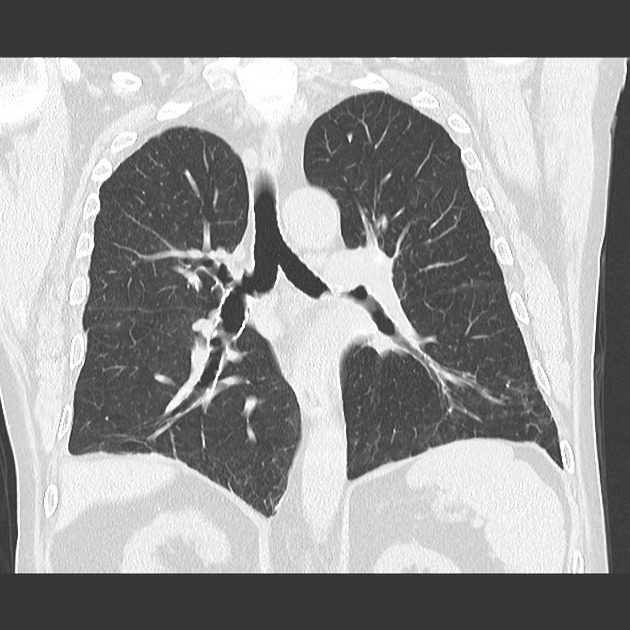
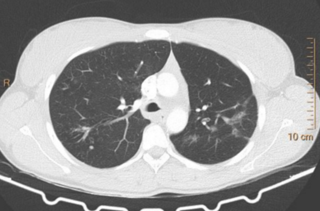
Bronchial stenosis, or bronchial strictures, are descriptive terms to denote regions of focal narrowing involving the bronchi. They can arise from a wide variety of etiologies.
On this page:
Images:
Pathology
Etiology
It can arise from a large range of etiological factors, which include:
-
non-malignant growths
-
infectious causes
-
mycobacterial
-
bacterial
respiratory (tracheobronchial) diphtheria
-
viral
influenza 13
-
fungal
-
endemic mycoses
-
-
-
inflammatory and infiltrative causes
as an extra-intestinal manifestation of inflammatory bowel disease 8
-
congenital
-
congenital bronchial stenosis
most commonly due to extrinsic compression by anomalous vascular structures
rarely as a contiguous extension ("long segment") of congenital tracheal stenosis 17
-
-
other causes
-
post-surgical
post lung transplantation
-
post-traumatic 11
delayed complication of tracheobronchial injury 11
-
corrosive injury
sequela of caustic (e.g. acid, alkali) aspiration
-
especially ferrous sulfate (iron) tablets
prolonged endobronchial intubation
-
ADVERTISEMENT: Supporters see fewer/no ads
Radiographic features
CT
Allows direction visualization of the segments of narrowing (MinIP images may be additionally helpful). May also show resorptive (obstructive) atelectasis of segments of lung distal to the stenosis.
See also
References
- 1. G Ferretti, F B Jouvan, F Thony, C Pison, M Coulomb. Benign noninflammatory bronchial stenosis: treatment with balloon dilation. (1995) Radiology. 196 (3): 831-4. doi:10.1148/radiology.196.3.7644651 - Pubmed
- 2. Choe K et.al . Tuberculous Bronchial Stenosis: CT Findings in 28 Cases. (1990) AJR. American journal of roentgenology. doi:10.2214/ajr.155.5.2120966 - Pubmed
- 3. Chung M et.al Bronchial Stenosis Due to Anthracofibrosis. (1998) Chest. doi:10.1378/chest.113.2.344 - Pubmed
- 4. Obusez EC, Jamjoom L, Kirsch J, Gildea T, Mohammed TL. Computed tomography correlation of airway disease with bronchoscopy: part I--nonneoplastic large airway diseases. (2014) Current problems in diagnostic radiology. 43 (5): 268-77. doi:10.1067/j.cpradiol.2014.05.002 - Pubmed
- 5. Grenier PA, Beigelman-Aubry C, Brillet PY. Nonneoplastic tracheal and bronchial stenoses. (2009) Radiologic clinics of North America. 47 (2): 243-60. doi:10.1016/j.rcl.2008.11.011 - Pubmed
- 6. Ingegnoli A, Corsi A, Verardo E, De Filippo M, Sverzellati N, Zompatori M. Uncommon causes of tracheobronchial stenosis and wall thickening: MDCT imaging. (2007) La Radiologia medica. 112 (8): 1132-41. doi:10.1007/s11547-007-0211-z - Pubmed
- 7. W. De Wever, V. Vandecaveye, S. Lanciotti, J.A. Verschakelen. Multidetector CT-generated virtual bronchoscopy: an illustrated review of the potential clinical indications. (2004) European Respiratory Journal. 23 (5): 776. doi:10.1183/09031936.04.00099804 - Pubmed
- 8. Papanikolaou I, Kagouridis K, Papiris SA. Patterns of airway involvement in inflammatory bowel diseases. (2014) World journal of gastrointestinal pathophysiology. 5 (4): 560-9. doi:10.4291/wjgp.v5.i4.560 - Pubmed
- 9. Baughman RP, Lower EE, Tami T. Upper airway. 4: Sarcoidosis of the upper respiratory tract (SURT). (2010) Thorax. 65 (2): 181-6. doi:10.1136/thx.2008.112896 - Pubmed
- 10. Kwak J, Koo G, Chung S et al. A Case of Significant Endobronchial Injury Due to Recurrent Iron Pill Aspiration. Tuberc Respir Dis (Seoul). 2015;78(4):440-4. doi:10.4046/trd.2015.78.4.440 - Pubmed
- 11. Bobocea A, Matache R, Codreşi M, Bolca C, Cordoş I. [Sleeve Resection of Right Main Bronchus for Posttraumatic Bronchial Stenosis]. Pneumologia. 2011;60(4):225-8. - Pubmed
- 12. Hadfield T, McEvoy P, Polotsky Y, Tzinserling V, Yakovlev A. The Pathology of Diphtheria. J INFECT DIS. 2000;181(s1):S116-20. doi:10.1086/315551 - Pubmed
- 13. Mailloux B, Burguete S, Stupka J, Sonetti D. Extensive Web-Like Endobronchial Membranous Stenosis in a Patient After H1N1 Influenza A Infectio. Chest. 2011;140(4):82A. doi:10.1378/chest.1117403
- 14. Barnes D, Gutiérrez Chacoff J, Benegas M et al. Central Airway Pathology: Clinic Features, CT Findings with Pathologic and Virtual Endoscopy Correlation. Insights Imaging. 2017;8(2):255-70. doi:10.1007/s13244-017-0545-6 - Pubmed
- 15. Schweiger C, Cohen A, Rutter M. Tracheal and Bronchial Stenoses and Other Obstructive Conditions. J Thorac Dis. 2016;8(11):3369-78. doi:10.21037/jtd.2016.11.74 - Pubmed
- 16. Smith M, Kou Y, Schweiger C, Lehenbauer D, de Alarcon A, Rutter M. Congenital Absence of Tracheal or Bronchial Rings. Otolaryngol Head Neck Surg. 2021;164(2):422-6. doi:10.1177/0194599820950725 - Pubmed
- 17. Fandiño M, Kozak F, Verchere C, Campbell A. Modified Slide Tracheoplasty in a Newborn with Bronchial and Carinal Stenosis. Int J Pediatr Otorhinolaryngol. 2013;77(12):2075-80. doi:10.1016/j.ijporl.2013.09.002 - Pubmed
Incoming Links
Related articles: Chest
- imaging techniques[+][+]
-
chest radiograph
- radiography
-
approach
- ABCDE
- ABCDEFGHI
- congenital heart disease
- medical devices in the thorax
- common lines and tubes
- nasogastric tubes
- endotracheal tubes
- central venous catheters
- esophageal temperature probe
- tracheostomy tube
- pleural catheters
- cardiac conduction devices
- prosthetic heart valve
- review areas
-
airspace opacification
- differential diagnoses of airspace opacification
- lobar consolidation
-
atelectasis
- mechanism-based
- morphology-based
- lobar lung collapse
- chest x-ray in the exam setting
- cardiomediastinal contour
- chest radiograph zones
- tracheal air column
- fissures
- normal chest x-ray appearance of the diaphragm
- nipple shadow
-
lines and stripes
- anterior junction line
- posterior junction line
- right paratracheal stripe
- left paratracheal stripe
- posterior tracheal stripe/tracheo-esophageal stripe
- posterior wall of bronchus intermedius
- right paraspinal line
- left paraspinal line
- aortic-pulmonary stripe
- aortopulmonary window
- azygo-esophageal recess
- spaces
- signs
- air bronchogram
- big rib sign
- Chang sign
- Chen sign
- coin lesion
- continuous diaphragm sign
- dense hilum sign
- double contour sign
- egg-on-a-string sign
- extrapleural sign
- finger in glove sign
- flat waist sign
- Fleischner sign
- ginkgo leaf sign
- Golden S sign
- Hampton hump
- haystack sign
- hilum convergence sign
- hilum overlay sign
- Hoffman-Rigler sign
- holly leaf sign
- incomplete border sign
- juxtaphrenic peak sign
- Kirklin sign
- medial stripe sign
- melting ice cube sign
- more black sign
- Naclerio V sign
- Palla sign
- pericardial fat tag sign
- Shmoo sign
- silhouette sign
- snowman sign
- spinnaker sign
- steeple sign
- straight left heart border sign
- third mogul sign
- tram-track sign
- walking man sign
- water bottle sign
- wave sign
- Westermark sign
- HRCT
-
chest radiograph
- airways
- bronchitis[+][+]
- small airways disease
-
bronchiectasis[+][+]
- broncho-arterial ratio
- related conditions
- differentials by distribution
- narrowing
-
tracheal stenosis[+][+]
- diffuse tracheal narrowing (differential)
-
bronchial stenosis
- diffuse airway narrowing (differential)
-
tracheal stenosis[+][+]
- diverticula[+][+]
- pulmonary edema[+][+]
-
interstitial lung disease (ILD)[+][+]
- Anti-Jo-1 antibody-positive interstitial lung disease
- drug-induced interstitial lung disease
-
hypersensitivity pneumonitis
- acute hypersensitivity pneumonitis
- subacute hypersensitivity pneumonitis
- chronic hypersensitivity pneumonitis
- etiology
- bird fancier's lung: pigeon fancier's lung
- farmer's lung
- cheese workers' lung
- bagassosis
- mushroom worker’s lung
- malt worker’s lung
- maple bark disease
- hot tub lung
- wine maker’s lung
- woodsman’s disease
- thatched roof lung
- tobacco grower’s lung
- potato riddler’s lung
- summer-type pneumonitis
- dry rot lung
- machine operator’s lung
- humidifier lung
- shower curtain disease
- furrier’s lung
- miller’s lung
- lycoperdonosis
- saxophone lung
-
idiopathic interstitial pneumonia (mnemonic)
- acute interstitial pneumonia (AIP)
- cryptogenic organizing pneumonia (COP)
- desquamative interstitial pneumonia (DIP)
- non-specific interstitial pneumonia (NSIP)
- idiopathic pleuroparenchymal fibroelastosis
- lymphoid interstitial pneumonia (LIP)
- respiratory bronchiolitis–associated interstitial lung disease (RB-ILD)
- usual interstitial pneumonia / idiopathic pulmonary fibrosis (UIP/IPF)
-
pneumoconioses
- fibrotic
- non-fibrotic
-
lung cancer[+][+]
-
non-small-cell lung cancer
-
adenocarcinoma
- pre-invasive tumors
- minimally invasive tumors
- invasive tumors
- variants of invasive carcinoma
- described imaging features
- adenosquamous carcinoma
- large cell carcinoma
- primary sarcomatoid carcinoma of the lung
- squamous cell carcinoma
- salivary gland-type tumors
-
adenocarcinoma
- pulmonary neuroendocrine tumors
- preinvasive lesions
-
lung cancer invasion patterns
- tumor spread through air spaces (STAS)
- presence of non-lepidic patterns such as acinar, papillary, solid, or micropapillary
- myofibroblastic stroma associated with invasive tumor cells
- pleural invasion
- vascular invasion
- tumors by location
- benign neoplasms
- pulmonary metastases
- lung cancer screening
- lung cancer staging
-
non-small-cell lung cancer


 Unable to process the form. Check for errors and try again.