
Palla sign
Citation, DOI, disclosures and article data
At the time the article was created Isaac Narouz had no recorded disclosures.
View Isaac Narouz's current disclosuresAt the time the article was last revised Craig Hacking had the following disclosures:
- Philips Australia, Paid speaker at Philips Spectral CT events (ongoing)
These were assessed during peer review and were determined to not be relevant to the changes that were made.
View Craig Hacking's current disclosures- Palla's sign
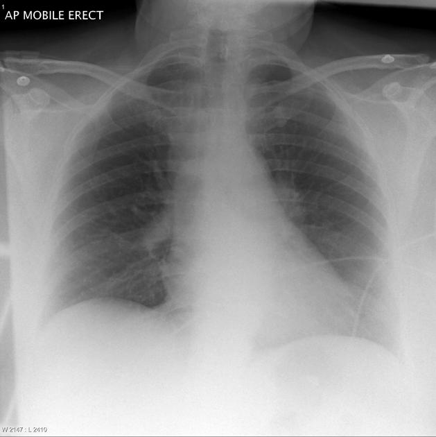
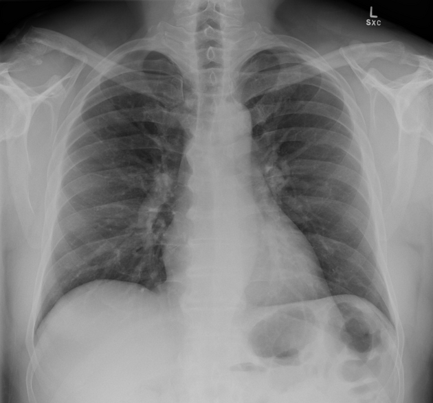
Palla sign is a sign seen on chest radiographs suggestive of pulmonary embolism, usually seen in the acute setting.
Although uncommon, it can be seen along with several other described signs of pulmonary embolus on chest radiography.
On this page:
Pathology
Palla sign describes an enlarged right descending pulmonary artery. When present with the Westermark sign, is suggestive of an occlusion of a lobar or segmental pulmonary artery, or widespread occlusion of small arteries.
Radiographic features
Plain radiograph
The descending interlobar branch of the right pulmonary artery is enlarged, causing a "sausage" appearance towards right middle lobe of the chest radiograph.
The sign has a low sensitivity and its specificity is not known.
History and etymology
The sign is named after Antonio Palla (1949-fl.2020), an Italian radiologist, who published his observations in 1983 that approximately 25% of patients with pulmonary embolism had an enlarged right descending pulmonary artery 1.
References
- 1. Palla A, Donnamaria V, Petruzzelli S, Rossi G, Riccetti G, Giuntini C. Enlargement of the right descending pulmonary artery in pulmonary embolism. AJR Am J Roentgenol. 1983 Sep;141(3):513-7.
- 2. Brenes-Salazar J A. Westermark's and Palla's signs in acute and chronic pulmonary embolism: Still valid in the current computed tomography era. J Emerg Trauma Shock. 2014 Jan-Mar; 7(1): 57–58.
- 3. Lee DS, Vo H, Franco A, Keshavamurthy J, Rotem E. Palla and Westermark Signs. J Thorac Imaging. 2017;32(4):7.
- 4. Ladeiras-Lopes R, Neto A, Costa C, et al. Hampton’s Hump and Palla’s Sign in Pulmonary Embolism. Circulation. 2013;127(18):1914-1915.
Incoming Links
Related articles: Chest
- imaging techniques
-
chest radiograph
- radiography[+][+]
-
approach
- ABCDE
- ABCDEFGHI
- congenital heart disease
- medical devices in the thorax
- common lines and tubes[+][+]
- nasogastric tubes
- endotracheal tubes
- central venous catheters
- esophageal temperature probe
- tracheostomy tube
- pleural catheters
- cardiac conduction devices
- prosthetic heart valve
- review areas
-
airspace opacification[+][+]
- differential diagnoses of airspace opacification
- lobar consolidation
-
atelectasis
- mechanism-based
- morphology-based
- lobar lung collapse
- chest x-ray in the exam setting[+][+]
- cardiomediastinal contour[+][+]
- chest radiograph zones[+][+]
- tracheal air column[+][+]
- fissures[+][+]
- normal chest x-ray appearance of the diaphragm[+][+]
- nipple shadow[+][+]
-
lines and stripes[+][+]
- anterior junction line
- posterior junction line
- right paratracheal stripe
- left paratracheal stripe
- posterior tracheal stripe/tracheo-esophageal stripe
- posterior wall of bronchus intermedius
- right paraspinal line
- left paraspinal line
- aortic-pulmonary stripe
- aortopulmonary window
- azygo-esophageal recess
- spaces[+][+]
- signs
- air bronchogram
- big rib sign
- Chang sign
- Chen sign
- coin lesion
- continuous diaphragm sign
- dense hilum sign
- double contour sign
- egg-on-a-string sign
- extrapleural sign
- finger in glove sign
- flat waist sign
- Fleischner sign
- ginkgo leaf sign
- Golden S sign
- Hampton hump
- haystack sign
- hilum convergence sign
- hilum overlay sign
- Hoffman-Rigler sign
- holly leaf sign
- incomplete border sign
- juxtaphrenic peak sign
- Kirklin sign
- medial stripe sign
- melting ice cube sign
- more black sign
- Naclerio V sign
- Palla sign
- pericardial fat tag sign
- Shmoo sign
- silhouette sign[+][+]
- snowman sign
- spinnaker sign
- steeple sign
- straight left heart border sign
- third mogul sign
- tram-track sign
- walking man sign
- water bottle sign
- wave sign
- Westermark sign
- HRCT[+][+]
-
chest radiograph
- airways[+][+]
- bronchitis
- small airways disease
-
bronchiectasis
- broncho-arterial ratio
- related conditions
- differentials by distribution
- narrowing
-
tracheal stenosis
- diffuse tracheal narrowing (differential)
-
bronchial stenosis
- diffuse airway narrowing (differential)
-
tracheal stenosis
- diverticula
- pulmonary edema[+][+]
-
interstitial lung disease (ILD)[+][+]
- Anti-Jo-1 antibody-positive interstitial lung disease
- drug-induced interstitial lung disease
-
hypersensitivity pneumonitis
- acute hypersensitivity pneumonitis
- subacute hypersensitivity pneumonitis
- chronic hypersensitivity pneumonitis
- etiology
- bird fancier's lung: pigeon fancier's lung
- farmer's lung
- cheese workers' lung
- bagassosis
- mushroom worker’s lung
- malt worker’s lung
- maple bark disease
- hot tub lung
- wine maker’s lung
- woodsman’s disease
- thatched roof lung
- tobacco grower’s lung
- potato riddler’s lung
- summer-type pneumonitis
- dry rot lung
- machine operator’s lung
- humidifier lung
- shower curtain disease
- furrier’s lung
- miller’s lung
- lycoperdonosis
- saxophone lung
-
idiopathic interstitial pneumonia (mnemonic)
- acute interstitial pneumonia (AIP)
- cryptogenic organizing pneumonia (COP)
- desquamative interstitial pneumonia (DIP)
- non-specific interstitial pneumonia (NSIP)
- idiopathic pleuroparenchymal fibroelastosis
- lymphoid interstitial pneumonia (LIP)
- respiratory bronchiolitis–associated interstitial lung disease (RB-ILD)
- usual interstitial pneumonia / idiopathic pulmonary fibrosis (UIP/IPF)
-
pneumoconioses
- fibrotic
- non-fibrotic
-
lung cancer[+][+]
-
non-small-cell lung cancer
-
adenocarcinoma
- pre-invasive tumors
- minimally invasive tumors
- invasive tumors
- variants of invasive carcinoma
- described imaging features
- adenosquamous carcinoma
- large cell carcinoma
- primary sarcomatoid carcinoma of the lung
- squamous cell carcinoma
- salivary gland-type tumors
-
adenocarcinoma
- pulmonary neuroendocrine tumors
- preinvasive lesions
-
lung cancer invasion patterns
- tumor spread through air spaces (STAS)
- presence of non-lepidic patterns such as acinar, papillary, solid, or micropapillary
- myofibroblastic stroma associated with invasive tumor cells
- pleural invasion
- vascular invasion
- tumors by location
- benign neoplasms
- pulmonary metastases
- lung cancer screening
- lung cancer staging
-
non-small-cell lung cancer


 Unable to process the form. Check for errors and try again.