
Nipple shadows
Citation, DOI, disclosures and article data
At the time the article was created Henry Knipe had no recorded disclosures.
View Henry Knipe's current disclosuresAt the time the article was last revised Liz Silverstone had no financial relationships to ineligible companies to disclose.
View Liz Silverstone's current disclosures- Nipple shadow
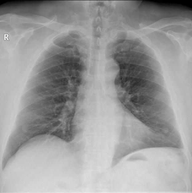
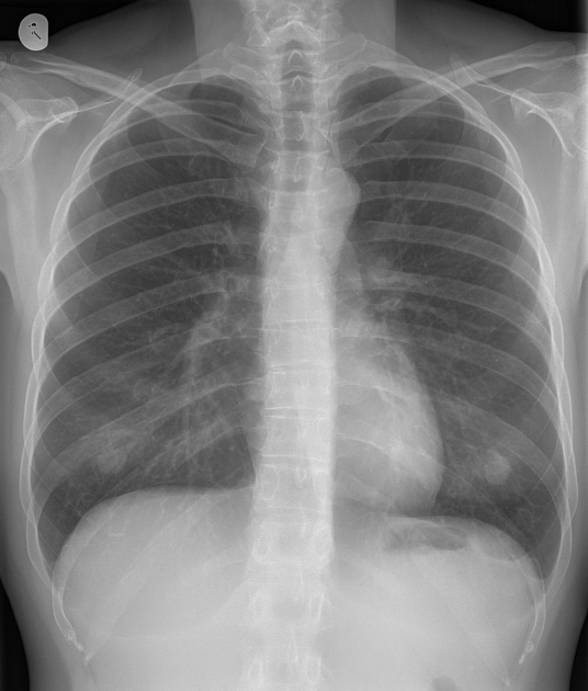
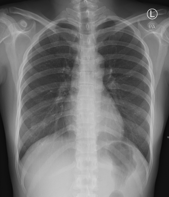
Nipple shadows refer to the silhouettes of the nipples on frontal chest radiographs, which may mimic solitary pulmonary nodules (SPNs).
On this page:
Epidemiology
Nipple shadows are apparent on ~7.5% (range 3.5-11%) of frontal chest x-rays 1.
Pathology
It has been proposed by Miller et al. that solitary pulmonary nodules that reach some or all of the following criteria be considered nipple shadows 2:
- bilateral and symmetric
- "fuzzy" margins or radiolucent "halo"
- sharp lateral border and poorly defined medial border (may be present only on PA projections 3)
- nodules are in a characteristic position:
- male: between the 5th and 6th ribs anteriorly
- female: at the inferior aspect of the breast shadow
- were not present on a very recent film
- prominent nipples may be visible on a lateral projection
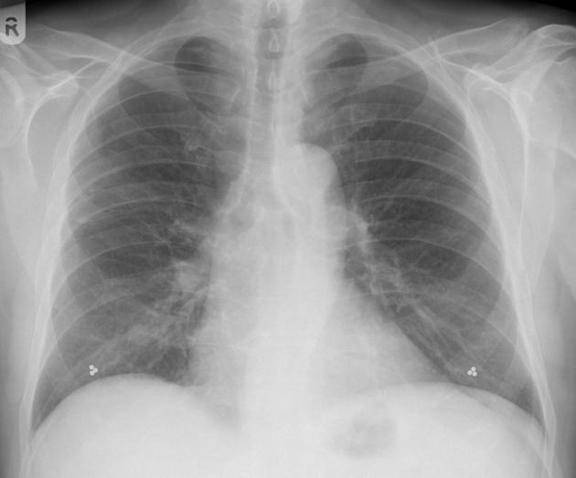
However, if there is doubt whether a nodular opacity represents a nipple shadow or not, a repeat chest x-ray with nipple markers should be performed 4, albeit at a financial cost and further radiation dose to the patient.
There is always the rare occurrence of a real solitary pulmonary nodule being overshadowed by a nipple shadow.
Radiographic features
Plain radiograph
On chest radiograph the following features are present:
- nipple shadows are nodular opacities that are consistent in shape, size and position 1,2:
- oval or round
- 5-15 mm in diameter
- between the 9th and 10th ribs posteriorly or the 5th and 6th ribs anteriorly
- tend to have incomplete margins due to their attachment to the chest wall 4
Differential diagnosis
- solitary pulmonary nodule
- pleural or extrapleural lesions 3
References
- 1. Ohwada A, Sato K, Tamori Y et-al. Visible male nipple shadows in chest radiographs. Respirology. 2005;10 (1): 111-5. doi:10.1111/j.1440-1843.2005.00653.x - Pubmed citation
- 2. Miller WT, Aronchick JM, Epstein DM et-al. The troublesome nipple shadow. AJR Am J Roentgenol. 1985;145 (3): 521-3. doi:10.2214/ajr.145.3.521 - Pubmed citation
- 3. Ferris RA, White AF. The round nipple shadow. Radiology. 1976;121 (2): 293-4. Pubmed citation
- 4. Reed JC. Chest Radiology. Mosby. ISBN:1437723454. Read it at Google Books - Find it at Amazon
- 5. Polnitsky CA, Toffler RB, Sherter CB. Nipple shadows in X-ray films. The New England journal of medicine. 305 (16): 956-7. doi:10.1056/NEJM198110153051616 - Pubmed
- 6. Moss ML. More on nipple shadows in x-ray films. The New England journal of medicine. 306 (3): 176. Pubmed
Incoming Links
- Pulmonary pseudonodules
- Bilateral nipple densities confirmed with nipple markers
- Nipple shadows
- Nipple shadows
- Male breast cancer
- Milk of calcium within breast cysts
- Solitary pulmonary nodule and nipple shadow
- Right-sided pneumonectomy
- Nipple shadows
- Subcutaneous chest wall lesion
- Bilateral nipple shadows
Related articles: Chest
- imaging techniques
-
chest radiograph
- radiography[+][+]
-
approach
- ABCDE
- ABCDEFGHI
- congenital heart disease
- medical devices in the thorax
- common lines and tubes[+][+]
- nasogastric tubes
- endotracheal tubes
- central venous catheters
- esophageal temperature probe
- tracheostomy tube
- pleural catheters
- cardiac conduction devices
- prosthetic heart valve
- review areas
-
airspace opacification[+][+]
- differential diagnoses of airspace opacification
- lobar consolidation
-
atelectasis
- mechanism-based
- morphology-based
- lobar lung collapse
- chest x-ray in the exam setting[+][+]
- cardiomediastinal contour[+][+]
- chest radiograph zones[+][+]
- tracheal air column[+][+]
- fissures[+][+]
- normal chest x-ray appearance of the diaphragm[+][+]
- nipple shadow
-
lines and stripes[+][+]
- anterior junction line
- posterior junction line
- right paratracheal stripe
- left paratracheal stripe
- posterior tracheal stripe/tracheo-esophageal stripe
- posterior wall of bronchus intermedius
- right paraspinal line
- left paraspinal line
- aortic-pulmonary stripe
- aortopulmonary window
- azygo-esophageal recess
- spaces[+][+]
- signs[+][+]
- air bronchogram
- big rib sign
- Chang sign
- Chen sign
- coin lesion
- continuous diaphragm sign
- dense hilum sign
- double contour sign
- egg-on-a-string sign
- extrapleural sign
- finger in glove sign
- flat waist sign
- Fleischner sign
- ginkgo leaf sign
- Golden S sign
- Hampton hump
- haystack sign
- hilum convergence sign
- hilum overlay sign
- Hoffman-Rigler sign
- holly leaf sign
- incomplete border sign
- juxtaphrenic peak sign
- Kirklin sign
- medial stripe sign
- melting ice cube sign
- more black sign
- Naclerio V sign
- Palla sign
- pericardial fat tag sign
- Shmoo sign
- silhouette sign
- snowman sign
- spinnaker sign
- steeple sign
- straight left heart border sign
- third mogul sign
- tram-track sign
- walking man sign
- water bottle sign
- wave sign
- Westermark sign
- HRCT[+][+]
-
chest radiograph
- airways[+][+]
- bronchitis
- small airways disease
-
bronchiectasis
- broncho-arterial ratio
- related conditions
- differentials by distribution
- narrowing
-
tracheal stenosis
- diffuse tracheal narrowing (differential)
-
bronchial stenosis
- diffuse airway narrowing (differential)
-
tracheal stenosis
- diverticula
- pulmonary edema[+][+]
-
interstitial lung disease (ILD)[+][+]
- Anti-Jo-1 antibody-positive interstitial lung disease
- drug-induced interstitial lung disease
-
hypersensitivity pneumonitis
- acute hypersensitivity pneumonitis
- subacute hypersensitivity pneumonitis
- chronic hypersensitivity pneumonitis
- etiology
- bird fancier's lung: pigeon fancier's lung
- farmer's lung
- cheese workers' lung
- bagassosis
- mushroom worker’s lung
- malt worker’s lung
- maple bark disease
- hot tub lung
- wine maker’s lung
- woodsman’s disease
- thatched roof lung
- tobacco grower’s lung
- potato riddler’s lung
- summer-type pneumonitis
- dry rot lung
- machine operator’s lung
- humidifier lung
- shower curtain disease
- furrier’s lung
- miller’s lung
- lycoperdonosis
- saxophone lung
-
idiopathic interstitial pneumonia (mnemonic)
- acute interstitial pneumonia (AIP)
- cryptogenic organizing pneumonia (COP)
- desquamative interstitial pneumonia (DIP)
- non-specific interstitial pneumonia (NSIP)
- idiopathic pleuroparenchymal fibroelastosis
- lymphoid interstitial pneumonia (LIP)
- respiratory bronchiolitis–associated interstitial lung disease (RB-ILD)
- usual interstitial pneumonia / idiopathic pulmonary fibrosis (UIP/IPF)
-
pneumoconioses
- fibrotic
- non-fibrotic
-
lung cancer[+][+]
-
non-small-cell lung cancer
-
adenocarcinoma
- pre-invasive tumors
- minimally invasive tumors
- invasive tumors
- variants of invasive carcinoma
- described imaging features
- adenosquamous carcinoma
- large cell carcinoma
- primary sarcomatoid carcinoma of the lung
- squamous cell carcinoma
- salivary gland-type tumors
-
adenocarcinoma
- pulmonary neuroendocrine tumors
- preinvasive lesions
-
lung cancer invasion patterns
- tumor spread through air spaces (STAS)
- presence of non-lepidic patterns such as acinar, papillary, solid, or micropapillary
- myofibroblastic stroma associated with invasive tumor cells
- pleural invasion
- vascular invasion
- tumors by location
- benign neoplasms
- pulmonary metastases
- lung cancer screening
- lung cancer staging
-
non-small-cell lung cancer


 Unable to process the form. Check for errors and try again.