
Peripherally inserted central catheter
Citation, DOI, disclosures and article data
At the time the article was created Jeremy Jones had no recorded disclosures.
View Jeremy Jones's current disclosuresAt the time the article was last revised Daniel MacManus had no financial relationships to ineligible companies to disclose.
View Daniel MacManus's current disclosures- Peripherally inserted central catheters
- PICC lines
- Peripherally inserted central catheter
- Peripherally inserted central catheters (PICCs)
- PICC
- PICCs
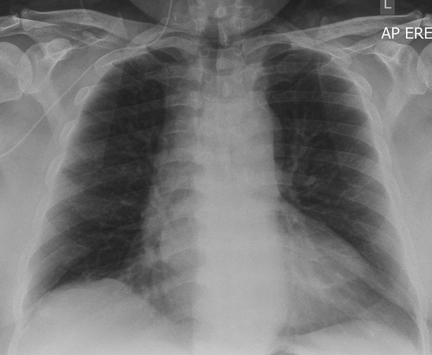
Peripherally inserted central catheters (PICCs), often incorrectly tautologically termed PICC lines, are a type of central venous catheter predominantly used amongst oncology patients and those with chronic diseases (e.g. cystic fibrosis).
They offer the ability to have long-term central venous access without the need to have a surgically or radiologically-inserted tunneled central venous catheter (e.g. Hickman catheter) or chest/brachial port.
On this page:
Indications
central venous access required for long-term IV administration of medication (e.g. chemotherapy or antibiotics) or parenteral nutrition
peripheral access when standard IV cannulation is difficult or impossible
Technique
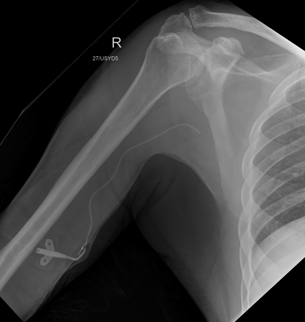
basilic and brachial veins are most commonly used; preprocedure ultrasound can be performed to identify an appropriately-sized vessel and ensure it is clot-free
sterile preparation and drape; ensure tourniquet is tight
subcutaneous infiltration of local anesthetic (e.g. lidocaine)
ultrasound-guided venous puncture followed by guidewire insertion; release the tourniquet
fluoroscopy to ensure guidewire position is venous
small skin incision at the puncture site
exchange puncture needle for peel-away sheath
removal of guidewire and insertion of PICC under fluoroscopic guidance to ensure tip is in an appropriate position (varies from institution to institution)
removal of the peel-away sheath
flush, secure and dress PICC
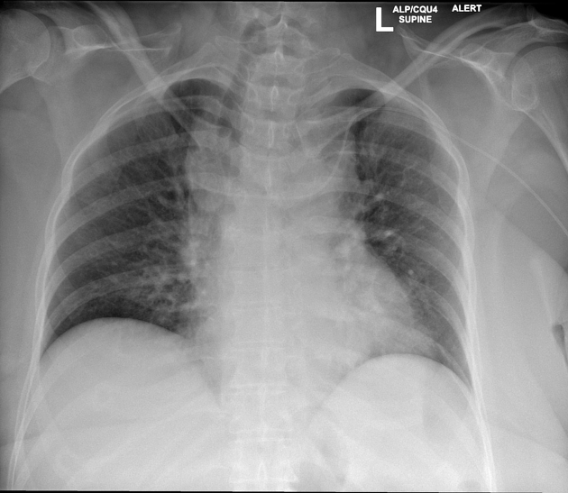
fluoroscopic spot acquisition/chest x-ray in inspiration to document correct position
ADVERTISEMENT: Supporters see fewer/no ads
Complications
Periprocedural
hemorrhage
arrhythmia
arterial puncture: less common with image guidance
malposition: less common with image guidance. Beware of anatomical variants such as a left SVC
allergic reaction 2
Late
line fracture/embolization or accidental withdrawal
formation of fibrin sheath
infection, e.g. catheter- or wound-related (most common) 3
tip migration
Quiz questions

References
- 1. Amerasekera SS, Jones CM, Patel R et-al. Imaging of the complications of peripherally inserted central venous catheters. Clin Radiol. 2009;64 (8): 832-40. doi:10.1016/j.crad.2009.02.021 - Pubmed citation
- 2. Funaki B. Central venous access: a primer for the diagnostic radiologist. AJR Am J Roentgenol. 2002;179 (2): 309-18. doi:10.2214/ajr.179.2.1790309 - Pubmed citation
- 3. Kim HJ, Yun J, Kim HJ et-al. Safety and effectiveness of central venous catheterization in patients with cancer: prospective observational study. J. Korean Med. Sci. 2010;25 (12): 1748-53. doi:10.3346/jkms.2010.25.12.1748 - Free text at pubmed - Pubmed citation
Incoming Links
- Medical devices in the head and neck
- Deep vein thrombosis
- Medical devices in the thorax
- Inferior cavoatrial junction
- Chest x-ray: PICC position (summary)
- Periprocedural anticoagulation
- Medical devices in the limbs
- Brachioradial artery
- Medical abbreviations and acronyms (P)
- Implantable port
- Central venous catheter
- Malpositioned PICC line secondary to retrosternal goitre
- Midline catheter
- Retained PICC line anchor securement system
- Extravascular migration of PICC line
- Surfactant deficiency disorder and thermal wrap
- Fractured peripherally inserted central catheter
- Surfactant deficiency disorder and Broviac line
- Malpositioned PICC and necrotising enterocolitis (NEC)
- Upper limb DVT - PICC line associated thrombus
- Dislodgement of PICC after contrast injection
- Dislodgement of PICC after contrast injection
- Malpositioned PICC
- Misplaced endotracheal tube
- Respiratory distress syndrome with pneumothorax
- Wolman disease
- Pulmonary embolisation of PICC fibrin sheath
- Right upper lobe atelectasis - intubated neonate
- Pediatric inflammatory multisystem syndrome (PIMS) post COVID-19
- Neonatal respiratory distress syndrome, pneumothorax, and PICC line malposition
- Incorrect peripherally inserted central catheter position
Related articles: Chest
- imaging techniques
-
chest radiograph
- radiography[+][+]
-
approach
- ABCDE
- ABCDEFGHI
- congenital heart disease
- medical devices in the thorax
- common lines and tubes
- nasogastric tubes[+][+]
- endotracheal tubes[+][+]
-
central venous catheters
- jugular venous catheter[+][+]
- peripherally inserted central catheters
- differential of left paramediastinal catheter positions
- esophageal temperature probe
- tracheostomy tube
- pleural catheters
- cardiac conduction devices
- prosthetic heart valve
- review areas
-
airspace opacification[+][+]
- differential diagnoses of airspace opacification
- lobar consolidation
-
atelectasis
- mechanism-based
- morphology-based
- lobar lung collapse
- chest x-ray in the exam setting[+][+]
- cardiomediastinal contour[+][+]
- chest radiograph zones[+][+]
- tracheal air column[+][+]
- fissures[+][+]
- normal chest x-ray appearance of the diaphragm[+][+]
- nipple shadow[+][+]
-
lines and stripes[+][+]
- anterior junction line
- posterior junction line
- right paratracheal stripe
- left paratracheal stripe
- posterior tracheal stripe/tracheo-esophageal stripe
- posterior wall of bronchus intermedius
- right paraspinal line
- left paraspinal line
- aortic-pulmonary stripe
- aortopulmonary window
- azygo-esophageal recess
- spaces[+][+]
- signs[+][+]
- air bronchogram
- big rib sign
- Chang sign
- Chen sign
- coin lesion
- continuous diaphragm sign
- dense hilum sign
- double contour sign
- egg-on-a-string sign
- extrapleural sign
- finger in glove sign
- flat waist sign
- Fleischner sign
- ginkgo leaf sign
- Golden S sign
- Hampton hump
- haystack sign
- hilum convergence sign
- hilum overlay sign
- Hoffman-Rigler sign
- holly leaf sign
- incomplete border sign
- juxtaphrenic peak sign
- Kirklin sign
- medial stripe sign
- melting ice cube sign
- more black sign
- Naclerio V sign
- Palla sign
- pericardial fat tag sign
- Shmoo sign
- silhouette sign
- snowman sign
- spinnaker sign
- steeple sign
- straight left heart border sign
- third mogul sign
- tram-track sign
- walking man sign
- water bottle sign
- wave sign
- Westermark sign
- HRCT[+][+]
-
chest radiograph
- airways[+][+]
- bronchitis
- small airways disease
-
bronchiectasis
- broncho-arterial ratio
- related conditions
- differentials by distribution
- narrowing
-
tracheal stenosis
- diffuse tracheal narrowing (differential)
-
bronchial stenosis
- diffuse airway narrowing (differential)
-
tracheal stenosis
- diverticula
- pulmonary edema[+][+]
-
interstitial lung disease (ILD)[+][+]
- Anti-Jo-1 antibody-positive interstitial lung disease
- drug-induced interstitial lung disease
-
hypersensitivity pneumonitis
- acute hypersensitivity pneumonitis
- subacute hypersensitivity pneumonitis
- chronic hypersensitivity pneumonitis
- etiology
- bird fancier's lung: pigeon fancier's lung
- farmer's lung
- cheese workers' lung
- bagassosis
- mushroom worker’s lung
- malt worker’s lung
- maple bark disease
- hot tub lung
- wine maker’s lung
- woodsman’s disease
- thatched roof lung
- tobacco grower’s lung
- potato riddler’s lung
- summer-type pneumonitis
- dry rot lung
- machine operator’s lung
- humidifier lung
- shower curtain disease
- furrier’s lung
- miller’s lung
- lycoperdonosis
- saxophone lung
-
idiopathic interstitial pneumonia (mnemonic)
- acute interstitial pneumonia (AIP)
- cryptogenic organizing pneumonia (COP)
- desquamative interstitial pneumonia (DIP)
- non-specific interstitial pneumonia (NSIP)
- idiopathic pleuroparenchymal fibroelastosis
- lymphoid interstitial pneumonia (LIP)
- respiratory bronchiolitis–associated interstitial lung disease (RB-ILD)
- usual interstitial pneumonia / idiopathic pulmonary fibrosis (UIP/IPF)
-
pneumoconioses
- fibrotic
- non-fibrotic
-
lung cancer[+][+]
-
non-small-cell lung cancer
-
adenocarcinoma
- pre-invasive tumors
- minimally invasive tumors
- invasive tumors
- variants of invasive carcinoma
- described imaging features
- adenosquamous carcinoma
- large cell carcinoma
- primary sarcomatoid carcinoma of the lung
- squamous cell carcinoma
- salivary gland-type tumors
-
adenocarcinoma
- pulmonary neuroendocrine tumors
- preinvasive lesions
-
lung cancer invasion patterns
- tumor spread through air spaces (STAS)
- presence of non-lepidic patterns such as acinar, papillary, solid, or micropapillary
- myofibroblastic stroma associated with invasive tumor cells
- pleural invasion
- vascular invasion
- tumors by location
- benign neoplasms
- pulmonary metastases
- lung cancer screening
- lung cancer staging
-
non-small-cell lung cancer


 Unable to process the form. Check for errors and try again.