
The thymus (plural: thymi) is a lymphoid organ in the anterior mediastinum responsible for the production and maturation of T-cells until puberty. It is a vital component of the immune system and plays a role in prevention of cancer 12, infection and autoimmunity 11.
On this page:
Gross anatomy
It is relatively large in infancy (weighing 25 g at birth) and grows considerably immediately after birth. Maximum weight of around 35g is achieved at puberty after which time it undergoes involution with progressive fatty replacement, reaching a weight of around 15g at 60 years of age. Involution occurs more rapidly in males. There can be a wide variation in size between patients 3.
The two lobes are usually asymmetric in size, the left lobe being higher and larger than the right 10. The lobes are occasionally united or may be separated by an intermediate lobe. Depending on age and size, the normal gland may extend from the lower border of the thyroid gland to the fourth costal cartilage.
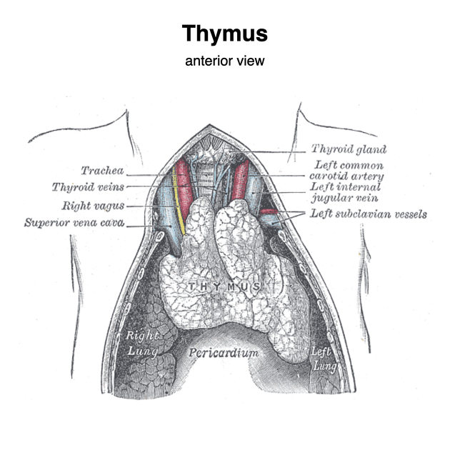
Relations
anteriorly: sternum, origins of the inferior strap muscles
posteriorly: pericardium, aortic arch, great vessels, left brachiocephalic vein, trachea
laterally: pleura, pretracheal fascia
Arterial supply
Venous drainage
Lymphatic drainage
parasternal, brachiocephalic and tracheobronchial lymph nodes
Innervation
sympathetic fibers entering with blood vessels that are vasomotor
Histology
The thymus is of a pinkish-grey color, soft, and lobulated on its surfaces.
Development
Embryologically it is derived from the third pharyngeal pouch. The thymus is the first of the lymphoid organs to be formed. Considerable growth occurs immediately after birth in response to antigen stimulation and demand for mature T cells. Genetic factors also influence dependence upon thymus immunological function. After fibrofatty atrophy, the thymus can grow back at any time in life, particularly after periods of stress.
Variant anatomy
variable location: ectopic and/or accessory thymic tissue may be located anywhere along the path of descent of the thymopharyngeal ducts, e.g. retrocaval, cervical, posterior mediastinal
variable shape: e.g. unilobed, trilobed, X-shaped, inverted V-shaped, etc.
Radiographic features
Plain radiograph
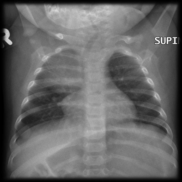
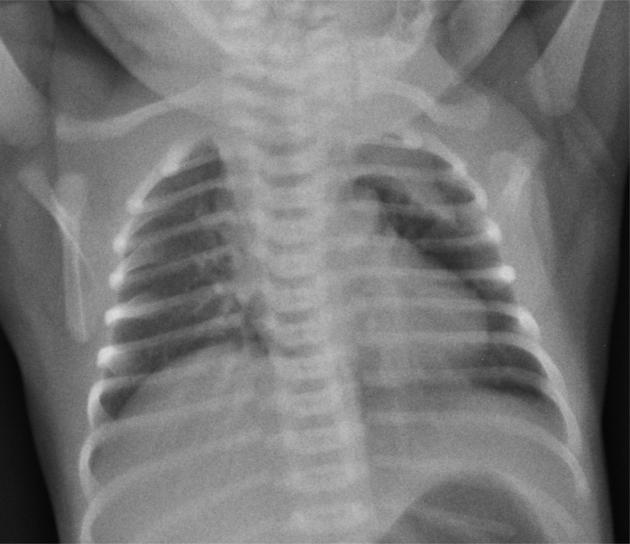
The thymus can be seen on chest radiographs within 24 hours after birth, then becomes smaller after the age of 2 years. It is rarely seen after the age of 8 years 10.
The thymus is seen as a triangular sail (thymic sail sign) frequently towards the right of the mediastinum. It has no mass effect on vascular structures or airways. The size can vary with inspiration.
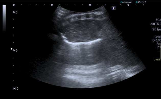
Ultrasound
typically relatively homogeneous background echogenicity similar to or slightly less than that of the liver and spleen, with scattered hyperechoic foci resembling a starry sky
its shape can be affected/distorted by cardiac pulsations and respiratory motion as it is soft and pliable and should not compress or displace adjacent structures
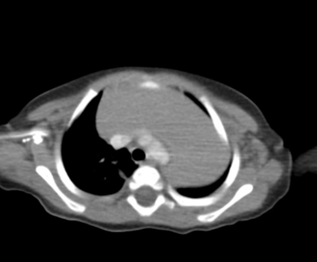
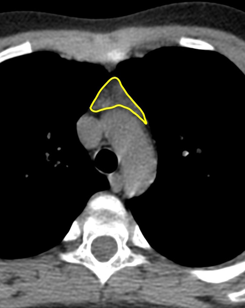
CT
typically uniformly soft tissue density, approximately 80 HU and isoattenuating to surrounding muscle
smooth outline with convex borders in childhood
triangular in adulthood, usually measures less than 1 cm in diameter 10
small blood vessels may be seen traversing it
MRI
typically demonstrates chemical shift artifact between in and out of phase images
-
differentiating normal from hyperplastic thymus can be difficult and guidelines for making this distinction and verifying the presence of normal thymus include 5:
absence of rounded soft-tissue masses >7 mm
absence of a convex contour of the thymus >19 years of age
absence of soft-tissue lobulation
absence of excessive thymic thickness (should be ≤1.3 cm when >20 years of age)
absence of a diagnosis associated with thymic enlargement or hyperplasia, e.g. myasthenia gravis
History and etymology
"Thymus" ultimately derives from the Greek word for the plant "thyme" θύμος ("to offer/sacrifice"), presumably because the plant was burnt on altars. Galen thought the thymus gland looked like a "warty excrescence" and resembled a bundle of plants 7.
The first good description of the thymus gland was recorded by Berengarius in 1524.
Related pathology
-
the normal thymus can mimic several pathologies:


 Unable to process the form. Check for errors and try again.